 Movie
Movie Controller

[English] 日本語
 Yorodumi
- PDB-3wc2: Crystal structure of C. albicans tRNA(His) guanylyltransferase (T... -
+ Open data

- Basic information
| Entry | Database: PDB / ID: 3wc2 | ||||||
|---|---|---|---|---|---|---|---|
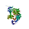
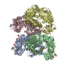
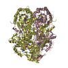
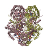
| Title | Crystal structure of C. albicans tRNA(His) guanylyltransferase (Thg1) with a tRNA(Phe)(GUG) | ||||||
 Components Components |
| ||||||
 Keywords Keywords | TRANSFERASE/RNA / TRANSFERASE-RNA complex | ||||||
| Function / homology | tRNA(His) guanylyltransferase (Thg1) / Alpha-Beta Plaits / 2-Layer Sandwich / Alpha Beta /  RNA / RNA (> 10) / : RNA / RNA (> 10) / :  Function and homology information Function and homology information | ||||||
| Biological species |  Candida albicans (yeast) Candida albicans (yeast) | ||||||
| Method | X-RAY DIFFRACTION / SYNCHROTRON / MOLECULAR REPLACEMENT / Resolution: 3.641 Å | ||||||
 Authors Authors | Nakamura, A. / Nemoto, T. / Sonoda, T. / Yamashita, K. / Tanaka, I. / Yao, M. | ||||||
 Citation Citation | Journal: Proc.Natl.Acad.Sci.USA / Year: 2013 Title: Structural basis of reverse nucleotide polymerization Authors: Nakamura, A. / Nemoto, T. / Heinemann, I.U. / Yamashita, K. / Sonoda, T. / Komoda, K. / Tanaka, I. / Soll, D. / Yao, M. | ||||||
| History |
|
- Structure visualization
| Structure viewer | Molecule: MolmilJmol/JSmol |
|---|
- Downloads & links
-Download
| PDBx/mmCIF format | 3wc2.cif.gz | 568.6 KB | Display | PDBx/mmCIF format |
|---|---|---|---|---|
| PDB format | pdb3wc2.ent.gz | 470.9 KB | Display | PDB format |
| PDBx/mmJSON format | 3wc2.json.gz | Tree view | PDBx/mmJSON format | |
| Others |  Other downloads Other downloads |
-Validation report
| Arichive directory | https://data.pdbj.org/pub/pdb/validation_reports/wc/3wc2ftp://data.pdbj.org/pub/pdb/validation_reports/wc/3wc2 | HTTPS FTP |
|---|
-Related structure data
| Related structure data |  3wbzSC  3wc0C  3wc1C S: Starting model for refinement C: citing same article ( |
|---|---|
| Similar structure data |

-Links
 PDBj
PDBj


- Assembly
| Deposited unit | 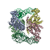
| ||||||||
|---|---|---|---|---|---|---|---|---|---|
| 1 |
| ||||||||
| Unit cell |
|
-Components
| #1: Protein | Mass: 32689.248 Da / Num. of mol.: 4 Source method: isolated from a genetically manipulated source Source: (gene. exp.) Candida albicans (yeast) / Strain: SC5314 / ATCC MYA-2876 / Gene: CaO19.7063, CAWG_05412, orf19.7063, THG1 / Plasmid: pET26b / Production host:  Escherichia coli (E. coli) / Strain (production host): B834(DE3) / References: UniProt: Q5AFK5 Escherichia coli (E. coli) / Strain (production host): B834(DE3) / References: UniProt: Q5AFK5#2: RNA chain | Mass: 24511.531 Da / Num. of mol.: 2 / Source method: obtained synthetically / Details: in vitro transcription |
|---|
-Experimental details
-Experiment
| Experiment | Method: X-RAY DIFFRACTION / Number of used crystals: 1 |
|---|
- Sample preparation
| Crystal | Density Matthews: 2.25 Å3/Da / Density % sol: 45.32 % |
|---|---|
| Crystal grow | Temperature: 293 K / Method: vapor diffusion, sitting drop / pH: 6 Details: Bis-Tris propan, CaCl2, PEG 3350, pH 6.0, vapor diffusion, sitting drop, temperature 293K |
-Data collection
| Diffraction | Mean temperature: 100 K | ||||||||||||||||||||||||||||||||||||||||||||||||||||||||||||||||||||||||||||||||||||||||||||||||||||
|---|---|---|---|---|---|---|---|---|---|---|---|---|---|---|---|---|---|---|---|---|---|---|---|---|---|---|---|---|---|---|---|---|---|---|---|---|---|---|---|---|---|---|---|---|---|---|---|---|---|---|---|---|---|---|---|---|---|---|---|---|---|---|---|---|---|---|---|---|---|---|---|---|---|---|---|---|---|---|---|---|---|---|---|---|---|---|---|---|---|---|---|---|---|---|---|---|---|---|---|---|---|
| Diffraction source | Source: SYNCHROTRON / Site: Photon Factory  / Beamline: BL-1A / Wavelength: 1.1 Å / Beamline: BL-1A / Wavelength: 1.1 Å | ||||||||||||||||||||||||||||||||||||||||||||||||||||||||||||||||||||||||||||||||||||||||||||||||||||
| Detector | Type: DECTRIS PILATUS 2M-F / Detector: PIXEL / Date: Jun 15, 2012 | ||||||||||||||||||||||||||||||||||||||||||||||||||||||||||||||||||||||||||||||||||||||||||||||||||||
| Radiation | Monochromator: Si(111) / Protocol: SINGLE WAVELENGTH / Monochromatic (M) / Laue (L): M / Scattering type: x-ray | ||||||||||||||||||||||||||||||||||||||||||||||||||||||||||||||||||||||||||||||||||||||||||||||||||||
| Radiation wavelength | Wavelength: 1.1 Å / Relative weight: 1 | ||||||||||||||||||||||||||||||||||||||||||||||||||||||||||||||||||||||||||||||||||||||||||||||||||||
| Reflection | Resolution: 3.64→48.711 Å / Num. obs: 18933 / % possible obs: 99.6 % / Observed criterion σ(I): -3 / Biso Wilson estimate: 125.88 Å2 / Rmerge(I) obs: 0.107 / Rrim(I) all: 0.113 / Net I/σ(I): 16.68 | ||||||||||||||||||||||||||||||||||||||||||||||||||||||||||||||||||||||||||||||||||||||||||||||||||||
| Reflection shell | Diffraction-ID: 1
|
- Processing
| Software |
| |||||||||||||||||||||||||||||||||||||||||||||||||||||||||||||||||||||||||||||||||||||||||||||||||||||||||||||||||||||||||||||||||||||||||||||||||||||||||||||||||||||||||||||||
|---|---|---|---|---|---|---|---|---|---|---|---|---|---|---|---|---|---|---|---|---|---|---|---|---|---|---|---|---|---|---|---|---|---|---|---|---|---|---|---|---|---|---|---|---|---|---|---|---|---|---|---|---|---|---|---|---|---|---|---|---|---|---|---|---|---|---|---|---|---|---|---|---|---|---|---|---|---|---|---|---|---|---|---|---|---|---|---|---|---|---|---|---|---|---|---|---|---|---|---|---|---|---|---|---|---|---|---|---|---|---|---|---|---|---|---|---|---|---|---|---|---|---|---|---|---|---|---|---|---|---|---|---|---|---|---|---|---|---|---|---|---|---|---|---|---|---|---|---|---|---|---|---|---|---|---|---|---|---|---|---|---|---|---|---|---|---|---|---|---|---|---|---|---|---|---|---|
| Refinement | Method to determine structure: MOLECULAR REPLACEMENT Starting model: 3WBZ Resolution: 3.641→38.09 Å / Occupancy max: 1 / Occupancy min: 1 / FOM work R set: 0.7663 / SU ML: 0.53 / Phase error: 29.43
| |||||||||||||||||||||||||||||||||||||||||||||||||||||||||||||||||||||||||||||||||||||||||||||||||||||||||||||||||||||||||||||||||||||||||||||||||||||||||||||||||||||||||||||||
| Solvent computation | Shrinkage radii: 0.9 Å / VDW probe radii: 1.11 Å / Solvent model: FLAT BULK SOLVENT MODEL | |||||||||||||||||||||||||||||||||||||||||||||||||||||||||||||||||||||||||||||||||||||||||||||||||||||||||||||||||||||||||||||||||||||||||||||||||||||||||||||||||||||||||||||||
| Displacement parameters | Biso max: 280.99 Å2 / Biso mean: 104.9006 Å2 / Biso min: 14.86 Å2 | |||||||||||||||||||||||||||||||||||||||||||||||||||||||||||||||||||||||||||||||||||||||||||||||||||||||||||||||||||||||||||||||||||||||||||||||||||||||||||||||||||||||||||||||
| Refinement step | Cycle: LAST / Resolution: 3.641→38.09 Å
| |||||||||||||||||||||||||||||||||||||||||||||||||||||||||||||||||||||||||||||||||||||||||||||||||||||||||||||||||||||||||||||||||||||||||||||||||||||||||||||||||||||||||||||||
| Refine LS restraints |
| |||||||||||||||||||||||||||||||||||||||||||||||||||||||||||||||||||||||||||||||||||||||||||||||||||||||||||||||||||||||||||||||||||||||||||||||||||||||||||||||||||||||||||||||
| LS refinement shell | Refine-ID: X-RAY DIFFRACTION / Total num. of bins used: 10
| |||||||||||||||||||||||||||||||||||||||||||||||||||||||||||||||||||||||||||||||||||||||||||||||||||||||||||||||||||||||||||||||||||||||||||||||||||||||||||||||||||||||||||||||
| Refinement TLS params. | Method: refined / Refine-ID: X-RAY DIFFRACTION
| |||||||||||||||||||||||||||||||||||||||||||||||||||||||||||||||||||||||||||||||||||||||||||||||||||||||||||||||||||||||||||||||||||||||||||||||||||||||||||||||||||||||||||||||
| Refinement TLS group |
|
