 Movie
Movie Controller

[English] 日本語
 Yorodumi
- PDB-3w1s: Crystal structure of Saccharomyces cerevisiae Atg12-Atg5 conjugat... -
+ Open data

- Basic information
| Entry | Database: PDB / ID: 3w1s | ||||||
|---|---|---|---|---|---|---|---|
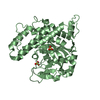
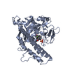
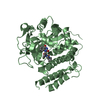
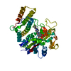
| Title | Crystal structure of Saccharomyces cerevisiae Atg12-Atg5 conjugate bound to the N-terminal domain of Atg16 | ||||||
 Components Components |
| ||||||
 Keywords Keywords |  LIGASE / Ubiquitin fold / E3-like / Atg3 binding / Isopeptide bond between Atg12 Gly186 and Atg5 Lys149 LIGASE / Ubiquitin fold / E3-like / Atg3 binding / Isopeptide bond between Atg12 Gly186 and Atg5 Lys149 | ||||||
| Function / homology |  Function and homology information Function and homology informationprotein lipidation involved in autophagosome assembly / cargo receptor ligand activity / Receptor Mediated Mitophagy / Atg8-family ligase activity / phagophore / Atg12-Atg5-Atg16 complex / : / C-terminal protein lipidation / vacuole-isolation membrane contact site / : ...protein lipidation involved in autophagosome assembly / cargo receptor ligand activity / Receptor Mediated Mitophagy / Atg8-family ligase activity / phagophore / Atg12-Atg5-Atg16 complex / : / C-terminal protein lipidation / vacuole-isolation membrane contact site / : / Macroautophagy / transferase complex / cytoplasm to vacuole targeting by the Cvt pathway / autophagosome organization / nucleophagy / piecemeal microautophagy of the nucleus / phagophore assembly site membrane / cellular response to nitrogen starvation / autophagy of mitochondrion / phagophore assembly site / autophagosome assembly / enzyme activator activity / autophagosome / macroautophagy / protein tag activity / autophagy / protein-macromolecule adaptor activity / protein transport / hydrolase activity / membrane / identical protein binding / nucleus / cytosol / cytoplasmSimilarity search - Function | ||||||
| Biological species |  Saccharomyces cerevisiae S288c (yeast) Saccharomyces cerevisiae S288c (yeast) | ||||||
| Method | X-RAY DIFFRACTION / SYNCHROTRON / MOLECULAR REPLACEMENT / Resolution: 2.6 Å | ||||||
 Authors Authors | Noda, N.N. / Fujioka, Y. / Hanada, T. / Ohsumi, Y. / Inagaki, F. | ||||||
 Citation Citation | Journal: Embo Rep. / Year: 2013 Title: Structure of the Atg12-Atg5 conjugate reveals a platform for stimulating Atg8-PE conjugation Authors: Noda, N.N. / Fujioka, Y. / Hanada, T. / Ohsumi, Y. / Inagaki, F. | ||||||
| History |
|
- Structure visualization
| Structure viewer | Molecule: MolmilJmol/JSmol |
|---|
- Downloads & links
-Download
| PDBx/mmCIF format | 3w1s.cif.gz | 86.3 KB | Display | PDBx/mmCIF format |
|---|---|---|---|---|
| PDB format | pdb3w1s.ent.gz | 64.1 KB | Display | PDB format |
| PDBx/mmJSON format | 3w1s.json.gz | Tree view | PDBx/mmJSON format | |
| Others |  Other downloads Other downloads |
-Validation report
| Arichive directory | https://data.pdbj.org/pub/pdb/validation_reports/w1/3w1sftp://data.pdbj.org/pub/pdb/validation_reports/w1/3w1s | HTTPS FTP |
|---|
-Related structure data


-Links
 PDBj
PDBj
- Assembly
| Deposited unit | 
| ||||||||
|---|---|---|---|---|---|---|---|---|---|
| 1 |
| ||||||||
| 2 | 
| ||||||||
| Unit cell |
|
-Components
| #1: Protein | Mass: 32620.820 Da / Num. of mol.: 1 / Fragment: UNP residues 1-284 Source method: isolated from a genetically manipulated source Source: (gene. exp.) Saccharomyces cerevisiae S288c (yeast) / Strain: ATCC 204508 / S288c / Gene: APG5, ATG5, Atg5(amino acids 1-284), P2601, YPL149W / Plasmid: pACYC184 / Production host:  Escherichia coli (E. coli) / Strain (production host): BL21(DE3) / References: UniProt: Q12380 Escherichia coli (E. coli) / Strain (production host): BL21(DE3) / References: UniProt: Q12380 |
|---|---|
| #2: Protein/peptide | / Cytoplasm to vacuole targeting protein 11 / SAP18 homolog Mass: 5792.501 Da / Num. of mol.: 1 / Fragment: UNP residues 1-46 Source method: isolated from a genetically manipulated source Source: (gene. exp.) Saccharomyces cerevisiae S288c (yeast) / Strain: ATCC 204508 / S288cGene: APG15, APG16, ATG16, Atg16(amino acids 1-46), CVT11, SAP18, YM8520.08C, YMR159C Plasmid: pGEX6p / Production host: Escherichia coli (E. coli) / Strain (production host): BL21(DE3) / References: UniProt: Q03818 |
| #3: Protein | Mass: 10341.172 Da / Num. of mol.: 1 / Fragment: UNP residues 100-186 Source method: isolated from a genetically manipulated source Source: (gene. exp.) Saccharomyces cerevisiae S288c (yeast) / Strain: ATCC 204508 / S288cGene: APG12, ATG12, Atg12(amino acids 100-186), YBR1506, YBR217W Plasmid: pACYC184 / Production host: Escherichia coli (E. coli) / Strain (production host): BL21(DE3) / References: UniProt: P38316 |
| #4: Water | ChemComp-HOH / Water Mass: 18.015 Da / Num. of mol.: 27 / Source method: isolated from a natural source / Formula: H2O Mass: 18.015 Da / Num. of mol.: 27 / Source method: isolated from a natural source / Formula: H2O |
-Experimental details
-Experiment
| Experiment | Method: X-RAY DIFFRACTION / Number of used crystals: 1 |
|---|
- Sample preparation
| Crystal | Density Matthews: 2.81 Å3/Da / Density % sol: 56.19 % |
|---|---|
| Crystal grow | Temperature: 293 K / Method: vapor diffusion, sitting drop / pH: 6.5 Details: 12% PEG10000, 0.5M potassium thiocyanate, 0.1M ADA, pH 6.5, VAPOR DIFFUSION, SITTING DROP, temperature 293K |
-Data collection
| Diffraction | Mean temperature: 95 K |
|---|---|
| Diffraction source | Source: SYNCHROTRON / Site: Photon Factory  / Beamline: AR-NW12A / Wavelength: 1 Å / Beamline: AR-NW12A / Wavelength: 1 Å |
| Detector | Type: ADSC QUANTUM 210 / Detector: CCD / Date: Jun 3, 2007 |
| Radiation | Protocol: SINGLE WAVELENGTH / Monochromatic (M) / Laue (L): M / Scattering type: x-ray |
| Radiation wavelength | Wavelength: 1 Å / Relative weight: 1 |
| Reflection | Resolution: 2.6→42.6 Å / Num. all: 17196 / Num. obs: 17173 / % possible obs: 99.9 % / Observed criterion σ(F): 0 / Observed criterion σ(I): -3 / Redundancy: 7.3 % / Biso Wilson estimate: 53.8 Å2 / Rsym value: 0.055 / Net I/σ(I): 9.6 |
| Reflection shell | Resolution: 2.6→2.69 Å / Redundancy: 7.4 % / Num. unique all: 1691 / Rsym value: 0.303 / % possible all: 100 |
- Processing
| Software |
| ||||||||||||||||||||||||||||||||||||||||||||||||||||||||||||||||||||||||||||||||
|---|---|---|---|---|---|---|---|---|---|---|---|---|---|---|---|---|---|---|---|---|---|---|---|---|---|---|---|---|---|---|---|---|---|---|---|---|---|---|---|---|---|---|---|---|---|---|---|---|---|---|---|---|---|---|---|---|---|---|---|---|---|---|---|---|---|---|---|---|---|---|---|---|---|---|---|---|---|---|---|---|---|
| Refinement | Method to determine structure: MOLECULAR REPLACEMENT Starting model: 2DYM, 1WZ3 Resolution: 2.6→42.6 Å / Rfactor Rfree error: 0.006 / Data cutoff high absF: 167335.04 / Data cutoff low absF: 0 / Isotropic thermal model: RESTRAINED / Cross valid method: THROUGHOUT / σ(F): 0 / Stereochemistry target values: Engh & Huber / Details: BULK SOLVENT MODEL USED
| ||||||||||||||||||||||||||||||||||||||||||||||||||||||||||||||||||||||||||||||||
| Solvent computation | Solvent model: FLAT MODEL / Bsol: 48.3649 Å2 / ksol: 0.38 e/Å3 | ||||||||||||||||||||||||||||||||||||||||||||||||||||||||||||||||||||||||||||||||
| Displacement parameters | Biso mean: 51.6 Å2
| ||||||||||||||||||||||||||||||||||||||||||||||||||||||||||||||||||||||||||||||||
| Refine analyze |
| ||||||||||||||||||||||||||||||||||||||||||||||||||||||||||||||||||||||||||||||||
| Refinement step | Cycle: LAST / Resolution: 2.6→42.6 Å
| ||||||||||||||||||||||||||||||||||||||||||||||||||||||||||||||||||||||||||||||||
| Refine LS restraints |
| ||||||||||||||||||||||||||||||||||||||||||||||||||||||||||||||||||||||||||||||||
| LS refinement shell | Resolution: 2.6→2.76 Å / Rfactor Rfree error: 0.022 / Total num. of bins used: 6
| ||||||||||||||||||||||||||||||||||||||||||||||||||||||||||||||||||||||||||||||||
| Xplor file |
|
