 Movie
Movie Controller

[English] 日本語
 Yorodumi
- PDB-3vf2: Crystal structure of the O-carbamoyltransferase TobZ M473I varian... -
+ Open data

- Basic information
| Entry | Database: PDB / ID: 3vf2 | ||||||
|---|---|---|---|---|---|---|---|
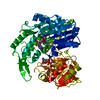
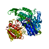
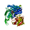
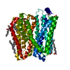
| Title | Crystal structure of the O-carbamoyltransferase TobZ M473I variant in complex with carbamoyl phosphate and ADP | ||||||
 Components Components | O-carbamoyltransferase TobZ | ||||||
 Keywords Keywords |  TRANSFERASE / antibiotic biosynthesis / substrate assisted catalysis / substrate channeling / adenylation / structural enzymology / enzyme evolution TRANSFERASE / antibiotic biosynthesis / substrate assisted catalysis / substrate channeling / adenylation / structural enzymology / enzyme evolution | ||||||
| Function / homology |  Function and homology information Function and homology informationnebramycin 5' synthase / tobramycin biosynthetic process / kanamycin biosynthetic process / carboxyl- or carbamoyltransferase activity / ligase activity / hydrolase activity / iron ion binding / ATP bindingSimilarity search - Function | ||||||
| Biological species |  Streptoalloteichus tenebrarius (bacteria) Streptoalloteichus tenebrarius (bacteria) | ||||||
| Method | X-RAY DIFFRACTION / MOLECULAR REPLACEMENT / Resolution: 2.9 Å | ||||||
 Authors Authors | Parthier, C. / Stubbs, M.T. / Goerlich, S. / Jaenecke, F. | ||||||
 Citation Citation | Journal: Angew.Chem.Int.Ed.Engl. / Year: 2012 Title: The O-Carbamoyltransferase TobZ Catalyzes an Ancient Enzymatic Reaction. Authors: Parthier, C. / Gorlich, S. / Jaenecke, F. / Breithaupt, C. / Brauer, U. / Fandrich, U. / Clausnitzer, D. / Wehmeier, U.F. / Bottcher, C. / Scheel, D. / Stubbs, M.T. | ||||||
| History |
|
- Structure visualization
| Structure viewer | Molecule: MolmilJmol/JSmol |
|---|
- Downloads & links
-Download
| PDBx/mmCIF format | 3vf2.cif.gz | 123.5 KB | Display | PDBx/mmCIF format |
|---|---|---|---|---|
| PDB format | pdb3vf2.ent.gz | 93.3 KB | Display | PDB format |
| PDBx/mmJSON format | 3vf2.json.gz | Tree view | PDBx/mmJSON format | |
| Others |  Other downloads Other downloads |
-Validation report
| Arichive directory | https://data.pdbj.org/pub/pdb/validation_reports/vf/3vf2ftp://data.pdbj.org/pub/pdb/validation_reports/vf/3vf2 | HTTPS FTP |
|---|
-Related structure data
| Related structure data |  3venSC  3veoC  3verC  3vesC 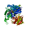 3vetC  3vewC  3vexC  3vezC  3vf4C  4e1bC S: Starting model for refinement C: citing same article ( |
|---|---|
| Similar structure data |


-Links
 PDBj
PDBj


- Assembly
| Deposited unit | 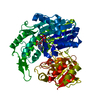
| ||||||||
|---|---|---|---|---|---|---|---|---|---|
| 1 |
| ||||||||
| Unit cell |
|
-Components
| #1: Protein | Mass: 63433.121 Da / Num. of mol.: 1 / Mutation: M473I Source method: isolated from a genetically manipulated source Source: (gene. exp.) Streptoalloteichus tenebrarius (bacteria)Strain: DSM40770T / Gene: tacA, tobZ / Plasmid: pUWL201PW / Production host: Streptomyces lividans (bacteria) / Strain (production host): TK24References: UniProt: Q70IY1, Transferases; Transferring one-carbon groups; Carboxy- and carbamoyltransferases |
|---|---|
| #2: Chemical | ChemComp-FE2 /   Mass: 55.845 Da / Num. of mol.: 1 / Source method: obtained synthetically / Formula: Fe Mass: 55.845 Da / Num. of mol.: 1 / Source method: obtained synthetically / Formula: Fe |
| #3: Chemical | ChemComp-K /   Mass: 39.098 Da / Num. of mol.: 1 / Source method: obtained synthetically / Formula: K Mass: 39.098 Da / Num. of mol.: 1 / Source method: obtained synthetically / Formula: K |
| #4: Chemical | ChemComp-CP / Carbamoyl phosphate  Mass: 141.020 Da / Num. of mol.: 1 / Source method: obtained synthetically / Formula: CH4NO5P Mass: 141.020 Da / Num. of mol.: 1 / Source method: obtained synthetically / Formula: CH4NO5P |
| #5: Chemical | ChemComp-ADP / Adenosine diphosphate  Mass: 427.201 Da / Num. of mol.: 1 / Source method: obtained synthetically / Formula: C10H15N5O10P2 / Comment: ADP, energy-carrying molecule*YM Mass: 427.201 Da / Num. of mol.: 1 / Source method: obtained synthetically / Formula: C10H15N5O10P2 / Comment: ADP, energy-carrying molecule*YM |
-Experimental details
-Experiment
| Experiment | Method: X-RAY DIFFRACTION / Number of used crystals: 1 |
|---|
- Sample preparation
| Crystal | Density Matthews: 3.14 Å3/Da / Density % sol: 60.86 % |
|---|---|
| Crystal grow | Temperature: 289 K / Method: vapor diffusion, hanging drop / pH: 6.5 Details: 1.6 M sodium potassium tartrate, 0.1 M MES, pH 6.5, VAPOR DIFFUSION, HANGING DROP, temperature 289K |
-Data collection
| Diffraction | Mean temperature: 100 K | ||||||||||||||||||||||||||||||||||||||||||||||||||||||||||||||||||||||||||||||||||||||||||||||||||
|---|---|---|---|---|---|---|---|---|---|---|---|---|---|---|---|---|---|---|---|---|---|---|---|---|---|---|---|---|---|---|---|---|---|---|---|---|---|---|---|---|---|---|---|---|---|---|---|---|---|---|---|---|---|---|---|---|---|---|---|---|---|---|---|---|---|---|---|---|---|---|---|---|---|---|---|---|---|---|---|---|---|---|---|---|---|---|---|---|---|---|---|---|---|---|---|---|---|---|---|
| Diffraction source | Source: ROTATING ANODE / Type: RIGAKU MICROMAX-007 / Wavelength: 1.5418 Å | ||||||||||||||||||||||||||||||||||||||||||||||||||||||||||||||||||||||||||||||||||||||||||||||||||
| Detector | Type: RIGAKU RAXIS IV++ / Detector: IMAGE PLATE / Date: Aug 24, 2011 | ||||||||||||||||||||||||||||||||||||||||||||||||||||||||||||||||||||||||||||||||||||||||||||||||||
| Radiation | Protocol: SINGLE WAVELENGTH / Monochromatic (M) / Laue (L): M / Scattering type: x-ray | ||||||||||||||||||||||||||||||||||||||||||||||||||||||||||||||||||||||||||||||||||||||||||||||||||
| Radiation wavelength | Wavelength: 1.5418 Å / Relative weight: 1 | ||||||||||||||||||||||||||||||||||||||||||||||||||||||||||||||||||||||||||||||||||||||||||||||||||
| Reflection | Resolution: 2.9→29.34 Å / Num. all: 19007 / Num. obs: 18717 / % possible obs: 98.5 % / Observed criterion σ(F): 0 / Observed criterion σ(I): -3 / Redundancy: 4.1 % / Biso Wilson estimate: 16.45 Å2 / Rmerge(I) obs: 0.148 / Net I/σ(I): 10.21 | ||||||||||||||||||||||||||||||||||||||||||||||||||||||||||||||||||||||||||||||||||||||||||||||||||
| Reflection shell |
|
- Processing
| Software |
| ||||||||||||||||||||||||||||||||||||||||||||||||||||||||
|---|---|---|---|---|---|---|---|---|---|---|---|---|---|---|---|---|---|---|---|---|---|---|---|---|---|---|---|---|---|---|---|---|---|---|---|---|---|---|---|---|---|---|---|---|---|---|---|---|---|---|---|---|---|---|---|---|---|
| Refinement | Method to determine structure: MOLECULAR REPLACEMENT Starting model: PDB ENTRY 3VEN Resolution: 2.9→29.336 Å / Occupancy max: 1 / Occupancy min: 1 / SU ML: 0.8 / Cross valid method: THROUGHOUT / σ(F): 1.36 / Phase error: 23.49 / Stereochemistry target values: ML
| ||||||||||||||||||||||||||||||||||||||||||||||||||||||||
| Solvent computation | Shrinkage radii: 0.83 Å / VDW probe radii: 1.1 Å / Solvent model: FLAT BULK SOLVENT MODEL / Bsol: 6.452 Å2 / ksol: 0.374 e/Å3 | ||||||||||||||||||||||||||||||||||||||||||||||||||||||||
| Displacement parameters | Biso max: 92.58 Å2 / Biso mean: 17.541 Å2 / Biso min: 3.41 Å2
| ||||||||||||||||||||||||||||||||||||||||||||||||||||||||
| Refinement step | Cycle: LAST / Resolution: 2.9→29.336 Å
| ||||||||||||||||||||||||||||||||||||||||||||||||||||||||
| Refine LS restraints |
| ||||||||||||||||||||||||||||||||||||||||||||||||||||||||
| LS refinement shell | Refine-ID: X-RAY DIFFRACTION / Total num. of bins used: 7
|
