 Movie
Movie Controller


+ Open data

- Basic information
| Entry | Database: PDB / ID: 3uxg | ||||||
|---|---|---|---|---|---|---|---|
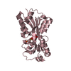
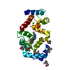
| Title | Crystal structure of RFXANK | ||||||
 Components Components |
| ||||||
 Keywords Keywords |  TRANSCRIPTION / histone / ankyrin repeat / structural genomics consortium / SGC TRANSCRIPTION / histone / ankyrin repeat / structural genomics consortium / SGC | ||||||
| Function / homology |  Function and homology information Function and homology informationRUNX2 regulates chondrocyte maturation / response to denervation involved in regulation of muscle adaptation / negative regulation of myotube differentiation / peptidyl-lysine deacetylation / positive regulation of protein sumoylation / negative regulation of transcription by competitive promoter binding / regulation of protein binding / protein deacetylation / cardiac muscle hypertrophy in response to stress / histone deacetylase ...RUNX2 regulates chondrocyte maturation / response to denervation involved in regulation of muscle adaptation / negative regulation of myotube differentiation / peptidyl-lysine deacetylation / positive regulation of protein sumoylation / negative regulation of transcription by competitive promoter binding / regulation of protein binding / protein deacetylation / cardiac muscle hypertrophy in response to stress / histone deacetylase / positive regulation of MHC class II biosynthetic process / protein lysine deacetylase activity / negative regulation of glycolytic process / SUMO transferase activity / histone deacetylase activity / negative regulation of gene expression, epigenetic / intercellular bridge / B cell activation / type I interferon-mediated signaling pathway / Notch-HLH transcription pathway / potassium ion binding / protein sumoylation / histone deacetylase complex / RUNX3 regulates p14-ARF / transcription repressor complex / SUMOylation of chromatin organization proteins / response to interleukin-1 / B cell differentiation / SUMOylation of intracellular receptors / negative regulation of DNA-binding transcription factor activity / NOTCH1 Intracellular Domain Regulates Transcription / Constitutive Signaling by NOTCH1 PEST Domain Mutants / Constitutive Signaling by NOTCH1 HD+PEST Domain Mutants / RNA polymerase II transcription regulator complex / histone deacetylase binding / positive regulation of DNA-binding transcription factor activity / nervous system development / DNA-binding transcription factor binding / Ras protein signal transduction / RNA polymerase II-specific DNA-binding transcription factor binding / molecular adaptor activity / nuclear speck / chromatin remodeling / inflammatory response / RNA polymerase II cis-regulatory region sequence-specific DNA binding / positive regulation of cell population proliferation / chromatin / regulation of transcription by RNA polymerase II / positive regulation of DNA-templated transcription / negative regulation of transcription by RNA polymerase II / positive regulation of transcription by RNA polymerase II / DNA binding / zinc ion binding / nucleoplasm / identical protein binding / nucleus / cytosol / cytoplasmSimilarity search - Function | ||||||
| Biological species |  Homo sapiens (human) Homo sapiens (human) | ||||||
| Method | X-RAY DIFFRACTION / SYNCHROTRON / MOLECULAR REPLACEMENT / molecular replacement / Resolution: 1.85 Å | ||||||
 Authors Authors | Tempel, W. / Chao, X. / Bian, C. / Li, Y. / Bountra, C. / Weigelt, J. / Arrowsmith, C.H. / Edwards, A.M. / Min, J. / Structural Genomics Consortium (SGC) | ||||||
 Citation Citation | Journal: Sci.Signal. / Year: 2012 Title: Sequence-Specific Recognition of a PxLPxI/L Motif by an Ankyrin Repeat Tumbler Lock. Authors: Xu, C. / Jin, J. / Bian, C. / Lam, R. / Tian, R. / Weist, R. / You, L. / Nie, J. / Bochkarev, A. / Tempel, W. / Tan, C.S. / Wasney, G.A. / Vedadi, M. / Gish, G.D. / Arrowsmith, C.H. / ...Authors: Xu, C. / Jin, J. / Bian, C. / Lam, R. / Tian, R. / Weist, R. / You, L. / Nie, J. / Bochkarev, A. / Tempel, W. / Tan, C.S. / Wasney, G.A. / Vedadi, M. / Gish, G.D. / Arrowsmith, C.H. / Pawson, T. / Yang, X.J. / Min, J. | ||||||
| History |
|
- Structure visualization
| Structure viewer | Molecule: MolmilJmol/JSmol |
|---|
- Downloads & links
-Download
| PDBx/mmCIF format | 3uxg.cif.gz | 89.1 KB | Display | PDBx/mmCIF format |
|---|---|---|---|---|
| PDB format | pdb3uxg.ent.gz | 66.7 KB | Display | PDB format |
| PDBx/mmJSON format | 3uxg.json.gz | Tree view | PDBx/mmJSON format | |
| Others |  Other downloads Other downloads |
-Validation report
| Arichive directory | https://data.pdbj.org/pub/pdb/validation_reports/ux/3uxgftp://data.pdbj.org/pub/pdb/validation_reports/ux/3uxg | HTTPS FTP |
|---|
-Related structure data
| Related structure data | 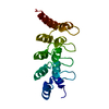 3so8SC 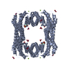 3uzdC 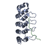 3v2oC 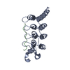 3v2xC 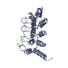 3v30C 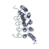 3v31C S: Starting model for refinement C: citing same article ( |
|---|---|
| Similar structure data |


-Links
 PDBj
PDBj


- Assembly
| Deposited unit | 
| ||||||||
|---|---|---|---|---|---|---|---|---|---|
| 1 |
| ||||||||
| 2 | 
| ||||||||
| Unit cell |
| ||||||||
| Components on special symmetry positions |
|
-Components
| #1: Protein | Mass: 18907.254 Da / Num. of mol.: 1 / Fragment: UNP residues 90-260 / Mutation: R199H Source method: isolated from a genetically manipulated source Source: (gene. exp.) Homo sapiens (human) / Gene: RFXANK, ANKRA1, RFXB / Plasmid: pET28-MHL / Production host:  Escherichia coli (E. coli) / Strain (production host): BL21-V2R-pRARE2 / References: UniProt: O14593 Escherichia coli (E. coli) / Strain (production host): BL21-V2R-pRARE2 / References: UniProt: O14593 | ||
|---|---|---|---|
| #2: Protein/peptide | / HD4 Mass: 1796.113 Da / Num. of mol.: 1 / Source method: obtained synthetically / Details: synthetic peptide / Source: (synth.) Homo sapiens (human) / References: UniProt: P56524, histone deacetylase | ||
| #3: Chemical | ChemComp-UNX /   Num. of mol.: 5 / Source method: obtained synthetically Num. of mol.: 5 / Source method: obtained synthetically#4: Water | ChemComp-HOH / | Water Mass: 18.015 Da / Num. of mol.: 117 / Source method: isolated from a natural source / Formula: H2O Mass: 18.015 Da / Num. of mol.: 117 / Source method: isolated from a natural source / Formula: H2O |
-Experimental details
-Experiment
| Experiment | Method: X-RAY DIFFRACTION / Number of used crystals: 1 |
|---|
- Sample preparation
| Crystal | Density Matthews: 2.7 Å3/Da / Density % sol: 54.5 % |
|---|---|
| Crystal grow | Temperature: 291 K / Method: vapor diffusion, sitting drop / pH: 7.5 Details: 25% PEG3350, 0.2M sodium chloride, 0.1M HEPES, pH 7.5, vapor diffusion, sitting drop, temperature 291K |
-Data collection
| Diffraction |
| |||||||||||||||||||||||||||||||||||||||||||||||||||||||||||||||||||||||||||||
|---|---|---|---|---|---|---|---|---|---|---|---|---|---|---|---|---|---|---|---|---|---|---|---|---|---|---|---|---|---|---|---|---|---|---|---|---|---|---|---|---|---|---|---|---|---|---|---|---|---|---|---|---|---|---|---|---|---|---|---|---|---|---|---|---|---|---|---|---|---|---|---|---|---|---|---|---|---|---|
| Diffraction source | Source: SYNCHROTRON / Site: APS  / Beamline: 19-ID / Wavelength: 0.97911 Å / Beamline: 19-ID / Wavelength: 0.97911 Å | |||||||||||||||||||||||||||||||||||||||||||||||||||||||||||||||||||||||||||||
| Detector | Type: ADSC QUANTUM 315 / Detector: CCD / Date: Jul 21, 2011 | |||||||||||||||||||||||||||||||||||||||||||||||||||||||||||||||||||||||||||||
| Radiation | Protocol: SINGLE WAVELENGTH / Monochromatic (M) / Laue (L): M / Scattering type: x-ray | |||||||||||||||||||||||||||||||||||||||||||||||||||||||||||||||||||||||||||||
| Radiation wavelength | Wavelength: 0.97911 Å / Relative weight: 1 | |||||||||||||||||||||||||||||||||||||||||||||||||||||||||||||||||||||||||||||
| Reflection | Resolution: 1.85→49.18 Å / Num. all: 18676 / Num. obs: 18636 / % possible obs: 99.96 % / Redundancy: 7.11 % / Rmerge(I) obs: 0.12 / Net I/σ(I): 15.0071 | |||||||||||||||||||||||||||||||||||||||||||||||||||||||||||||||||||||||||||||
| Reflection shell |
|
-Phasing
| Phasing | Method: molecular replacement |
|---|
- Processing
| Software |
| |||||||||||||||||||||||||||||||||||||||||||||||||||||||||||||||||||||||||||||||||||||||||||||||||||||||||||||||||||||||||||||||||||||||||||||||||||
|---|---|---|---|---|---|---|---|---|---|---|---|---|---|---|---|---|---|---|---|---|---|---|---|---|---|---|---|---|---|---|---|---|---|---|---|---|---|---|---|---|---|---|---|---|---|---|---|---|---|---|---|---|---|---|---|---|---|---|---|---|---|---|---|---|---|---|---|---|---|---|---|---|---|---|---|---|---|---|---|---|---|---|---|---|---|---|---|---|---|---|---|---|---|---|---|---|---|---|---|---|---|---|---|---|---|---|---|---|---|---|---|---|---|---|---|---|---|---|---|---|---|---|---|---|---|---|---|---|---|---|---|---|---|---|---|---|---|---|---|---|---|---|---|---|---|---|---|---|
| Refinement | Method to determine structure: MOLECULAR REPLACEMENT Starting model: pdb entry 3SO8 Resolution: 1.85→39.86 Å / Cor.coef. Fo:Fc: 0.946 / Cor.coef. Fo:Fc free: 0.92 / WRfactor Rfree: 0.206 / WRfactor Rwork: 0.172 / Occupancy max: 1 / Occupancy min: 0.5 / SU B: 5.718 / SU ML: 0.078 / Cross valid method: THROUGHOUT / σ(F): 0 / ESU R Free: 0.121 / Stereochemistry target values: MAXIMUM LIKELIHOOD Details: HYDROGENS HAVE BEEN ADDED IN THE RIDING POSITIONS U VALUES : WITH TLS ADDED. Arp/warp, coot and the molprobity server were also used during refinement. Residues 110 through 116 of chain A ...Details: HYDROGENS HAVE BEEN ADDED IN THE RIDING POSITIONS U VALUES : WITH TLS ADDED. Arp/warp, coot and the molprobity server were also used during refinement. Residues 110 through 116 of chain A are poorly defined by electron density.
| |||||||||||||||||||||||||||||||||||||||||||||||||||||||||||||||||||||||||||||||||||||||||||||||||||||||||||||||||||||||||||||||||||||||||||||||||||
| Solvent computation | Ion probe radii: 0.8 Å / Shrinkage radii: 0.8 Å / VDW probe radii: 1.4 Å / Solvent model: MASK | |||||||||||||||||||||||||||||||||||||||||||||||||||||||||||||||||||||||||||||||||||||||||||||||||||||||||||||||||||||||||||||||||||||||||||||||||||
| Displacement parameters | Biso max: 70.12 Å2 / Biso mean: 20.7861 Å2 / Biso min: 5.31 Å2
| |||||||||||||||||||||||||||||||||||||||||||||||||||||||||||||||||||||||||||||||||||||||||||||||||||||||||||||||||||||||||||||||||||||||||||||||||||
| Refinement step | Cycle: LAST / Resolution: 1.85→39.86 Å
| |||||||||||||||||||||||||||||||||||||||||||||||||||||||||||||||||||||||||||||||||||||||||||||||||||||||||||||||||||||||||||||||||||||||||||||||||||
| Refine LS restraints |
| |||||||||||||||||||||||||||||||||||||||||||||||||||||||||||||||||||||||||||||||||||||||||||||||||||||||||||||||||||||||||||||||||||||||||||||||||||
| LS refinement shell | Refine-ID: X-RAY DIFFRACTION / Total num. of bins used: 20
| |||||||||||||||||||||||||||||||||||||||||||||||||||||||||||||||||||||||||||||||||||||||||||||||||||||||||||||||||||||||||||||||||||||||||||||||||||
| Refinement TLS params. | Method: refined / Refine-ID: X-RAY DIFFRACTION
| |||||||||||||||||||||||||||||||||||||||||||||||||||||||||||||||||||||||||||||||||||||||||||||||||||||||||||||||||||||||||||||||||||||||||||||||||||
| Refinement TLS group |
|
