 Movie
Movie Controller

[English] 日本語
 Yorodumi
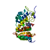
- PDB-3u4h: CD38 structure-based inhibitor design using the N1-cyclic inosine... -
+ Open data

- Basic information
| Entry | Database: PDB / ID: 3u4h | ||||||
|---|---|---|---|---|---|---|---|
| Title | CD38 structure-based inhibitor design using the N1-cyclic inosine 5'-diphosphate ribose template | ||||||
 Components Components | ADP-ribosyl cyclase 1 Cyclic ADP-ribose Cyclic ADP-ribose | ||||||
 Keywords Keywords | HYDROLASE/HYDROLASE INHIBITOR / non-hydrolyzable inhibitor / two domains / cADPR cyclization / hydrolysis / 8-amino-N1-cIDPR / HYDROLASE-HYDROLASE INHIBITOR complex | ||||||
| Function / homology |  Function and homology information Function and homology informationphosphorus-oxygen lyase activity / 2'-phospho-ADP-ribosyl cyclase/2'-phospho-cyclic-ADP-ribose transferase / artery smooth muscle contraction / Nicotinate metabolism / NAD+ nucleosidase activity / ADP-ribosyl cyclase/cyclic ADP-ribose hydrolase / NAD metabolic process / NAD+ nucleotidase, cyclic ADP-ribose generating / NADP+ nucleosidase activity / negative regulation of bone resorption ...phosphorus-oxygen lyase activity / 2'-phospho-ADP-ribosyl cyclase/2'-phospho-cyclic-ADP-ribose transferase / artery smooth muscle contraction / Nicotinate metabolism / NAD+ nucleosidase activity / ADP-ribosyl cyclase/cyclic ADP-ribose hydrolase / NAD metabolic process / NAD+ nucleotidase, cyclic ADP-ribose generating / NADP+ nucleosidase activity / negative regulation of bone resorption / response to hydroperoxide / long-term synaptic depression / Hydrolases; Glycosylases; Hydrolysing N-glycosyl compounds / B cell proliferation / response to retinoic acid / positive regulation of B cell proliferation / positive regulation of vasoconstriction / response to interleukin-1 / response to progesterone / female pregnancy / apoptotic signaling pathway / B cell receptor signaling pathway / positive regulation of insulin secretion / negative regulation of neuron projection development / response to estradiol / positive regulation of cytosolic calcium ion concentration / transferase activity / positive regulation of cell growth / basolateral plasma membrane / response to hypoxia / response to xenobiotic stimulus / negative regulation of DNA-templated transcription / negative regulation of apoptotic process / positive regulation of DNA-templated transcription / cell surface / signal transduction / extracellular exosome / membrane / identical protein binding / plasma membraneSimilarity search - Function | ||||||
| Biological species |  Homo sapiens (human) Homo sapiens (human) | ||||||
| Method | X-RAY DIFFRACTION / SYNCHROTRON / MOLECULAR REPLACEMENT / Resolution: 1.878 Å | ||||||
 Authors Authors | Liu, Q. / Hao, Q. / Lee, H.C. / Graeff, R. | ||||||
 Citation Citation | Journal: Plos One / Year: 2013 Title: CD38 Structure-Based Inhibitor Design Using the N1-Cyclic Inosine 5'-Diphosphate Ribose Template Authors: Moreau, C. / Liu, Q. / Graeff, R. / Wagner, G.K. / Thomas, M.P. / Swarbrick, J.M. / Shuto, S. / Lee, H.C. / Hao, Q. / Potter, B.V.L. | ||||||
| History |
|
- Structure visualization
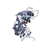
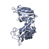
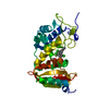
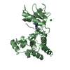
| Structure viewer | Molecule: MolmilJmol/JSmol |
|---|
- Downloads & links
-Download
| PDBx/mmCIF format | 3u4h.cif.gz | 224.6 KB | Display | PDBx/mmCIF format |
|---|---|---|---|---|
| PDB format | pdb3u4h.ent.gz | 179.4 KB | Display | PDB format |
| PDBx/mmJSON format | 3u4h.json.gz | Tree view | PDBx/mmJSON format | |
| Others |  Other downloads Other downloads |
-Validation report
| Arichive directory | https://data.pdbj.org/pub/pdb/validation_reports/u4/3u4hftp://data.pdbj.org/pub/pdb/validation_reports/u4/3u4h | HTTPS FTP |
|---|
-Related structure data
| Related structure data |  3u4iC  2pgjS C: citing same article ( S: Starting model for refinement |
|---|---|
| Similar structure data |


-Links
 PDBj
PDBj
- Assembly
| Deposited unit | 
| ||||||||
|---|---|---|---|---|---|---|---|---|---|
| 1 | 
| ||||||||
| 2 | 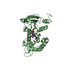
| ||||||||
| Unit cell |
|
-Components
| #1: Protein | Cyclic ADP-ribose / Cyclic ADP-ribose hydrolase 1 / cADPr hydrolase 1 / T10 Mass: 30380.393 Da / Num. of mol.: 2 / Fragment: extracellular domain, enzymatic domain / Mutation: Q49T, N100D, N164D, N209D, N219D Source method: isolated from a genetically manipulated source Source: (gene. exp.) Homo sapiens (human) / Gene: CD38 / Production host:  Pichia Pastoris (fungus) / References: UniProt: P28907, NAD+ glycohydrolase Pichia Pastoris (fungus) / References: UniProt: P28907, NAD+ glycohydrolase#2: Chemical |   Mass: 557.300 Da / Num. of mol.: 2 / Source method: obtained synthetically / Formula: C15H21N5O14P2 Mass: 557.300 Da / Num. of mol.: 2 / Source method: obtained synthetically / Formula: C15H21N5O14P2#3: Water | ChemComp-HOH / | Water Mass: 18.015 Da / Num. of mol.: 251 / Source method: isolated from a natural source / Formula: H2O Mass: 18.015 Da / Num. of mol.: 251 / Source method: isolated from a natural source / Formula: H2O |
|---|
-Experimental details
-Experiment
| Experiment | Method: X-RAY DIFFRACTION / Number of used crystals: 1 |
|---|
- Sample preparation
| Crystal | Density Matthews: 2.27 Å3/Da / Density % sol: 45.83 % |
|---|---|
| Crystal grow | Temperature: 298 K / Method: vapor diffusion, hanging drop / pH: 6 Details: 100mM MES, 12% PEG 4000, pH 6.0, VAPOR DIFFUSION, HANGING DROP, temperature 298K |
-Data collection
| Diffraction | Mean temperature: 100 K |
|---|---|
| Diffraction source | Source: SYNCHROTRON / Site: CHESS  / Beamline: A1 / Wavelength: 0.9778 Å / Beamline: A1 / Wavelength: 0.9778 Å |
| Detector | Type: ADSC QUANTUM 210 / Detector: CCD / Date: Oct 4, 2006 |
| Radiation | Monochromator: Si 111 CHANNEL / Protocol: SINGLE WAVELENGTH / Monochromatic (M) / Laue (L): M / Scattering type: x-ray |
| Radiation wavelength | Wavelength: 0.9778 Å / Relative weight: 1 |
| Reflection | Resolution: 1.87→50 Å / Num. all: 38912 / Num. obs: 38912 / % possible obs: 90.5 % / Observed criterion σ(F): 0 / Observed criterion σ(I): 0 |
| Reflection shell | Resolution: 1.87→1.94 Å / % possible all: 64.3 |
- Processing
| Software |
| ||||||||||||||||||||||||||||||||||||||||||||||||||||||||||||||||||||||||||||||||||||||||||||||||||||||||||||||||||||||||||||||||||||||||||||||||||||||||||||||||||||||||||||||||||||||||||||||||||||||||||||||||||||||||||||||||||||||||||||||||||||||||||||||||||||||||||||||||||||||||||||||||||||||||||||
|---|---|---|---|---|---|---|---|---|---|---|---|---|---|---|---|---|---|---|---|---|---|---|---|---|---|---|---|---|---|---|---|---|---|---|---|---|---|---|---|---|---|---|---|---|---|---|---|---|---|---|---|---|---|---|---|---|---|---|---|---|---|---|---|---|---|---|---|---|---|---|---|---|---|---|---|---|---|---|---|---|---|---|---|---|---|---|---|---|---|---|---|---|---|---|---|---|---|---|---|---|---|---|---|---|---|---|---|---|---|---|---|---|---|---|---|---|---|---|---|---|---|---|---|---|---|---|---|---|---|---|---|---|---|---|---|---|---|---|---|---|---|---|---|---|---|---|---|---|---|---|---|---|---|---|---|---|---|---|---|---|---|---|---|---|---|---|---|---|---|---|---|---|---|---|---|---|---|---|---|---|---|---|---|---|---|---|---|---|---|---|---|---|---|---|---|---|---|---|---|---|---|---|---|---|---|---|---|---|---|---|---|---|---|---|---|---|---|---|---|---|---|---|---|---|---|---|---|---|---|---|---|---|---|---|---|---|---|---|---|---|---|---|---|---|---|---|---|---|---|---|---|---|---|---|---|---|---|---|---|---|---|---|---|---|---|---|---|---|---|---|---|---|---|---|---|---|---|---|---|---|---|---|---|---|---|---|---|---|---|---|---|---|---|---|---|---|---|---|---|---|---|
| Refinement | Method to determine structure: MOLECULAR REPLACEMENT Starting model: 2PGJ Resolution: 1.878→25.246 Å / Occupancy max: 1 / Occupancy min: 1 / SU ML: 0.25 / σ(F): 1.97 / Phase error: 27.4 / Stereochemistry target values: ML
| ||||||||||||||||||||||||||||||||||||||||||||||||||||||||||||||||||||||||||||||||||||||||||||||||||||||||||||||||||||||||||||||||||||||||||||||||||||||||||||||||||||||||||||||||||||||||||||||||||||||||||||||||||||||||||||||||||||||||||||||||||||||||||||||||||||||||||||||||||||||||||||||||||||||||||||
| Solvent computation | Shrinkage radii: 0.9 Å / VDW probe radii: 1.11 Å / Solvent model: FLAT BULK SOLVENT MODEL / Bsol: 60.098 Å2 / ksol: 0.4 e/Å3 | ||||||||||||||||||||||||||||||||||||||||||||||||||||||||||||||||||||||||||||||||||||||||||||||||||||||||||||||||||||||||||||||||||||||||||||||||||||||||||||||||||||||||||||||||||||||||||||||||||||||||||||||||||||||||||||||||||||||||||||||||||||||||||||||||||||||||||||||||||||||||||||||||||||||||||||
| Displacement parameters | Biso max: 167.58 Å2 / Biso mean: 47.2435 Å2 / Biso min: 9.54 Å2
| ||||||||||||||||||||||||||||||||||||||||||||||||||||||||||||||||||||||||||||||||||||||||||||||||||||||||||||||||||||||||||||||||||||||||||||||||||||||||||||||||||||||||||||||||||||||||||||||||||||||||||||||||||||||||||||||||||||||||||||||||||||||||||||||||||||||||||||||||||||||||||||||||||||||||||||
| Refinement step | Cycle: LAST / Resolution: 1.878→25.246 Å
| ||||||||||||||||||||||||||||||||||||||||||||||||||||||||||||||||||||||||||||||||||||||||||||||||||||||||||||||||||||||||||||||||||||||||||||||||||||||||||||||||||||||||||||||||||||||||||||||||||||||||||||||||||||||||||||||||||||||||||||||||||||||||||||||||||||||||||||||||||||||||||||||||||||||||||||
| Refine LS restraints |
| ||||||||||||||||||||||||||||||||||||||||||||||||||||||||||||||||||||||||||||||||||||||||||||||||||||||||||||||||||||||||||||||||||||||||||||||||||||||||||||||||||||||||||||||||||||||||||||||||||||||||||||||||||||||||||||||||||||||||||||||||||||||||||||||||||||||||||||||||||||||||||||||||||||||||||||
| LS refinement shell | Refine-ID: X-RAY DIFFRACTION / Total num. of bins used: 14
| ||||||||||||||||||||||||||||||||||||||||||||||||||||||||||||||||||||||||||||||||||||||||||||||||||||||||||||||||||||||||||||||||||||||||||||||||||||||||||||||||||||||||||||||||||||||||||||||||||||||||||||||||||||||||||||||||||||||||||||||||||||||||||||||||||||||||||||||||||||||||||||||||||||||||||||
| Refinement TLS params. | Method: refined / Refine-ID: X-RAY DIFFRACTION
| ||||||||||||||||||||||||||||||||||||||||||||||||||||||||||||||||||||||||||||||||||||||||||||||||||||||||||||||||||||||||||||||||||||||||||||||||||||||||||||||||||||||||||||||||||||||||||||||||||||||||||||||||||||||||||||||||||||||||||||||||||||||||||||||||||||||||||||||||||||||||||||||||||||||||||||
| Refinement TLS group |
|
