 Movie
Movie Controller


+ Open data

- Basic information
| Entry | Database: PDB / ID: 3tz7 | ||||||
|---|---|---|---|---|---|---|---|
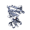
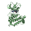
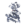
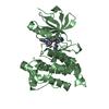
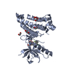
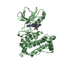
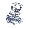
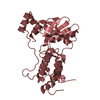
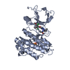
| Title | Kinase domain of cSrc in complex with RL103 | ||||||
 Components Components | Proto-oncogene tyrosine-protein kinase Src | ||||||
 Keywords Keywords | TRANSFERASE/TRANSFERASE INHIBITOR / Type II / Allosteric / DFG-out / Drug resistance mutations / Tyrosin-protein kinase / TRANSFERASE-TRANSFERASE INHIBITOR complex | ||||||
| Function / homology |  Function and homology information Function and homology informationSignaling by ERBB2 / Nuclear signaling by ERBB4 / PIP3 activates AKT signaling / Signaling by SCF-KIT / Regulation of KIT signaling / Signaling by EGFR / GAB1 signalosome / Regulation of gap junction activity / FCGR activation / PECAM1 interactions ...Signaling by ERBB2 / Nuclear signaling by ERBB4 / PIP3 activates AKT signaling / Signaling by SCF-KIT / Regulation of KIT signaling / Signaling by EGFR / GAB1 signalosome / Regulation of gap junction activity / FCGR activation / PECAM1 interactions / CD28 co-stimulation / CTLA4 inhibitory signaling / EPHA-mediated growth cone collapse / Ephrin signaling / G alpha (i) signalling events / GP1b-IX-V activation signalling / Recycling pathway of L1 / Thrombin signalling through proteinase activated receptors (PARs) / VEGFR2 mediated cell proliferation / RAF activation / PI5P, PP2A and IER3 Regulate PI3K/AKT Signaling / RET signaling / Receptor Mediated Mitophagy / ADP signalling through P2Y purinoceptor 1 / Downregulation of ERBB4 signaling / EPH-ephrin mediated repulsion of cells / Cyclin D associated events in G1 / Regulation of RUNX3 expression and activity / Activated NTRK3 signals through PI3K / Downstream signal transduction / MAP2K and MAPK activation / Integrin signaling / GRB2:SOS provides linkage to MAPK signaling for Integrins / MET activates PTK2 signaling / Extra-nuclear estrogen signaling / EPHB-mediated forward signaling / p130Cas linkage to MAPK signaling for integrins / VEGFA-VEGFR2 Pathway / connexin binding / osteoclast development / progesterone receptor signaling pathway / negative regulation of intrinsic apoptotic signaling pathway / bone resorption / extrinsic component of cytoplasmic side of plasma membrane / negative regulation of extrinsic apoptotic signaling pathway / non-specific protein-tyrosine kinase / non-membrane spanning protein tyrosine kinase activity / epidermal growth factor receptor signaling pathway / cell junction / protein phosphatase binding / protein tyrosine kinase activity / mitochondrial inner membrane / cell differentiation / cytoskeleton / endosome membrane / regulation of cell cycle / cell adhesion / cell cycle / phosphorylation / signaling receptor binding / focal adhesion / innate immune response / heme binding / perinuclear region of cytoplasm / protein-containing complex / ATP binding / membrane / nucleus / plasma membrane / cytosolSimilarity search - Function | ||||||
| Biological species |  Gallus gallus (chicken) Gallus gallus (chicken) | ||||||
| Method | X-RAY DIFFRACTION / MOLECULAR REPLACEMENT / molecular replacement / Resolution: 3.3 Å | ||||||
 Authors Authors | Gruetter, C. / Richters, A. / Rauh, D. | ||||||
 Citation Citation | Journal: To be Published Title: Overcoming Gatekeeper Mutations in cSrc and Abl by Hybrid Compound Design Authors: Richters, A. / Getlik, M. / Gruetter, C. / Schneider, R. / Heuckmann, J.M. / Heynck, S. / Sos, M.L. / Thomas, R.K. / Rauh, D. | ||||||
| History |
|
- Structure visualization
| Structure viewer | Molecule: MolmilJmol/JSmol |
|---|
- Downloads & links
-Download
| PDBx/mmCIF format | 3tz7.cif.gz | 117.8 KB | Display | PDBx/mmCIF format |
|---|---|---|---|---|
| PDB format | pdb3tz7.ent.gz | 90.6 KB | Display | PDB format |
| PDBx/mmJSON format | 3tz7.json.gz | Tree view | PDBx/mmJSON format | |
| Others |  Other downloads Other downloads |
-Validation report
| Arichive directory | https://data.pdbj.org/pub/pdb/validation_reports/tz/3tz7ftp://data.pdbj.org/pub/pdb/validation_reports/tz/3tz7 | HTTPS FTP |
|---|
-Related structure data
| Related structure data | 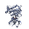 3tz8C  3tz9C  3f3vS S: Starting model for refinement C: citing same article ( |
|---|---|
| Similar structure data |


-Links
 PDBj
PDBj- Assembly
| Deposited unit | 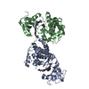
| ||||||||
|---|---|---|---|---|---|---|---|---|---|
| 1 | 
| ||||||||
| 2 | 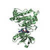
| ||||||||
| Unit cell |
|
-Components
| #1: Protein | / Proto-oncogene c-Src / pp60c-src / p60-Src Mass: 32742.711 Da / Num. of mol.: 2 / Fragment: Kinase Domain, UNP residues 251-533 / Mutation: S345C Source method: isolated from a genetically manipulated source Source: (gene. exp.) Gallus gallus (chicken) / Gene: SRC, v-Src sarcoma viral oncogene / Plasmid: pSKB3 / Production host:  Escherichia coli (E. coli) / Strain (production host): BL21(DE3) Escherichia coli (E. coli) / Strain (production host): BL21(DE3)References: UniProt: P00523, non-specific protein-tyrosine kinase#2: Chemical |   Mass: 620.747 Da / Num. of mol.: 2 / Source method: obtained synthetically / Formula: C34H40N10O2 Mass: 620.747 Da / Num. of mol.: 2 / Source method: obtained synthetically / Formula: C34H40N10O2 |
|---|
-Experimental details
-Experiment
| Experiment | Method: X-RAY DIFFRACTION / Number of used crystals: 1 |
|---|
- Sample preparation
| Crystal | Density Matthews: 3.09 Å3/Da / Density % sol: 60.2 % |
|---|---|
| Crystal grow | Temperature: 293 K / Method: vapor diffusion, hanging drop / pH: 6.5 Details: 10% PEG 20000, 15% glycerol, 100mM MES, pH 6.5, VAPOR DIFFUSION, HANGING DROP, temperature 293K |
-Data collection
| Diffraction | Mean temperature: 100 K | |||||||||||||||||||||||||||||||||||||||||||||||||||||||||||||||||||||||||||||||||||||||||||||||||||
|---|---|---|---|---|---|---|---|---|---|---|---|---|---|---|---|---|---|---|---|---|---|---|---|---|---|---|---|---|---|---|---|---|---|---|---|---|---|---|---|---|---|---|---|---|---|---|---|---|---|---|---|---|---|---|---|---|---|---|---|---|---|---|---|---|---|---|---|---|---|---|---|---|---|---|---|---|---|---|---|---|---|---|---|---|---|---|---|---|---|---|---|---|---|---|---|---|---|---|---|---|
| Diffraction source | Source: ROTATING ANODE / Type: BRUKER AXS MICROSTAR / Wavelength: 1.5417 Å | |||||||||||||||||||||||||||||||||||||||||||||||||||||||||||||||||||||||||||||||||||||||||||||||||||
| Detector | Type: MAR scanner 345 mm plate / Detector: IMAGE PLATE / Date: May 21, 2009 / Details: Osmic | |||||||||||||||||||||||||||||||||||||||||||||||||||||||||||||||||||||||||||||||||||||||||||||||||||
| Radiation | Monochromator: Multilayer Optic / Protocol: SINGLE WAVELENGTH / Monochromatic (M) / Laue (L): M / Scattering type: x-ray | |||||||||||||||||||||||||||||||||||||||||||||||||||||||||||||||||||||||||||||||||||||||||||||||||||
| Radiation wavelength | Wavelength: 1.5417 Å / Relative weight: 1 | |||||||||||||||||||||||||||||||||||||||||||||||||||||||||||||||||||||||||||||||||||||||||||||||||||
| Reflection | Resolution: 3.3→40 Å / Num. all: 11797 / Num. obs: 11298 / % possible obs: 95.8 % / Observed criterion σ(I): -3 / Redundancy: 2.21 % / Biso Wilson estimate: 18.547 Å2 / Rmerge(I) obs: 0.063 / Net I/σ(I): 15.24 | |||||||||||||||||||||||||||||||||||||||||||||||||||||||||||||||||||||||||||||||||||||||||||||||||||
| Reflection shell | Diffraction-ID: 1
|
-Phasing
| Phasing | Method: molecular replacement | |||||||||
|---|---|---|---|---|---|---|---|---|---|---|
| Phasing MR | Model details: Phaser MODE: MR_AUTO
|
- Processing
| Software |
| |||||||||||||||||||||||||||||||||||||||||||||||||||||||||||||||||
|---|---|---|---|---|---|---|---|---|---|---|---|---|---|---|---|---|---|---|---|---|---|---|---|---|---|---|---|---|---|---|---|---|---|---|---|---|---|---|---|---|---|---|---|---|---|---|---|---|---|---|---|---|---|---|---|---|---|---|---|---|---|---|---|---|---|---|
| Refinement | Method to determine structure: MOLECULAR REPLACEMENT Starting model: PDB ENTRY 3F3V Resolution: 3.3→28.58 Å / Cor.coef. Fo:Fc: 0.913 / Cor.coef. Fo:Fc free: 0.839 / WRfactor Rfree: 0.2198 / WRfactor Rwork: 0.1614 / Occupancy max: 1 / Occupancy min: 0.5 / FOM work R set: 0.8237 / SU B: 23.681 / SU ML: 0.397 / SU Rfree: 0.5484 / Cross valid method: THROUGHOUT / σ(F): 0 / ESU R Free: 0.548 / Stereochemistry target values: MAXIMUM LIKELIHOOD Details: HYDROGENS HAVE BEEN ADDED IN THE RIDING POSITIONS U VALUES: REFINED INDIVIDUALLY
| |||||||||||||||||||||||||||||||||||||||||||||||||||||||||||||||||
| Solvent computation | Ion probe radii: 0.8 Å / Shrinkage radii: 0.8 Å / VDW probe radii: 1.4 Å / Solvent model: MASK | |||||||||||||||||||||||||||||||||||||||||||||||||||||||||||||||||
| Displacement parameters | Biso max: 60.95 Å2 / Biso mean: 24.4479 Å2 / Biso min: 2 Å2
| |||||||||||||||||||||||||||||||||||||||||||||||||||||||||||||||||
| Refinement step | Cycle: LAST / Resolution: 3.3→28.58 Å
| |||||||||||||||||||||||||||||||||||||||||||||||||||||||||||||||||
| Refine LS restraints |
| |||||||||||||||||||||||||||||||||||||||||||||||||||||||||||||||||
| LS refinement shell | Resolution: 3.3→3.385 Å / Total num. of bins used: 20
|
