 Movie
Movie Controller

[English] 日本語
 Yorodumi
- PDB-3tmn: THE BINDING OF L-VALYL-L-TRYPTOPHAN TO CRYSTALLINE THERMOLYSIN IL... -
+ Open data

- Basic information
| Entry | Database: PDB / ID: 3tmn | |||||||||
|---|---|---|---|---|---|---|---|---|---|---|
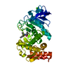
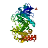
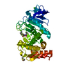
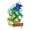
| Title | THE BINDING OF L-VALYL-L-TRYPTOPHAN TO CRYSTALLINE THERMOLYSIN ILLUSTRATES THE MODE OF INTERACTION OF A PRODUCT OF PEPTIDE HYDROLYSIS | |||||||||
 Components Components | THERMOLYSIN | |||||||||
 Keywords Keywords | HYDROLASE/HYDROLASE INHIBITOR / HYDROLASE / METALLOPROTEINASE / HYDROLASE-HYDROLASE INHIBITOR COMPLEX | |||||||||
| Function / homology |  Function and homology informationthermolysin / metalloendopeptidase activity / proteolysis / extracellular region / metal ion binding Function and homology informationthermolysin / metalloendopeptidase activity / proteolysis / extracellular region / metal ion bindingSimilarity search - Function | |||||||||
| Biological species |  | |||||||||
| Method | X-RAY DIFFRACTION / MOLECULAR SUPSTATUTION / Resolution: 1.7 Å | |||||||||
 Authors Authors | Holden, H.M. / Matthews, B.W. | |||||||||
 Citation Citation | Journal: J.Biol.Chem. / Year: 1988 Title: The binding of L-valyl-L-tryptophan to crystalline thermolysin illustrates the mode of interaction of a product of peptide hydrolysis. Authors: Holden, H.M. / Matthews, B.W. #1: Journal: Science / Year: 1987Title: Structures of Two Thermolysin-Inhibitor Complexes that Differ by a Single Hydrogen Bond Authors: Tronrud, D.E. / Holden, H.M. / Matthews, B.W. #2: Journal: Eur.J.Biochem. / Year: 1986Title: Crystallographic Structural Analysis of Phosphoramidates as Inhibitors and Transition-State Analogs of Thermolysin Authors: Tronrud, D.E. / Monzingo, A.F. / Matthews, B.W. #3: Journal: Biochemistry / Year: 1984Title: Binding of N-Carboxymethyl Dipepetide Inhibitors to Thermolysin Determined by X-Ray Crystallography. A Novel Class of Transition-State Analogues for Zinc Peptidases Authors: Monzingo, A.F. / Matthews, B.W. #4: Journal: Biochemistry / Year: 1984Title: An Interactive Computer Graphics Study of Thermolysin-Catalyzed Peptide Cleavage and Inhibition by N-Carboxymethyl Dipeptides Authors: Hangauer, D.G. / Monzingo, A.F. / Matthews, B.W. #5: Journal: Biochemistry / Year: 1983Title: Structural Analysis of the Inhibition of Thermolysin by an Active-Site-Directed Irreversible Inhibitor Authors: Holmes, M.A. / Tronrud, D.E. / Matthews, B.W. #6: Journal: Biochemistry / Year: 1982Title: Structure of a Mercaptan-Thermolysin Complex Illustrates Mode of Inhibition of Zinc Proteases by Substrate-Analogue Mercaptans Authors: Monzingo, A.F. / Matthews, B.W. #7: Journal: J.Mol.Biol. / Year: 1982Title: Structure of Thermolysin Refined at 1.6 Angstroms Resolution Authors: Holmes, M.A. / Matthews, B.W. #8: Journal: Biochemistry / Year: 1981Title: Binding of Hydroxamic Acid Inhibitors to Crystalline Thermolysin Suggests a Pentacoordinate Zinc Intermediate in Catalysis Authors: Holmes, M.A. / Matthews, B.W. #9: Journal: J.Biol.Chem. / Year: 1979Title: Binding of the Biproduct Analog L-Benzylsuccinic Acid to Thermolysin Determined by X-Ray Crystallography Authors: Bolognesi, M.C. / Matthews, B.W. #10: Journal: J.Biol.Chem. / Year: 1977Title: Comparison of the Structures of Carboxypeptidase a and Thermolysin Authors: Kester, W.R. / Matthews, B.W. #11: Journal: J.Mol.Biol. / Year: 1977Title: A Crystallographic Study of the Complex of Phosphoramidon with Thermolysin. A Model for the Presumed Catalytic Transition State and for the Binding of Extended Substrates Authors: Weaver, L.H. / Kester, W.R. / Matthews, B.W. #12: Journal: Biochemistry / Year: 1977Title: Crystallographic Study of the Binding of Dipeptide Inhibitors to Thermolysin. Implications for the Mechanism of Catalysis Authors: Kester, W.R. / Matthews, B.W. #13: Journal: Biochemistry / Year: 1976Title: Role of Calcium in the Thermal Stability of Thermolysin Authors: Dahlquist, F.W. / Long, J.W. / Bigbee, W.L. #14: Journal: Proc.Natl.Acad.Sci.USA / Year: 1975Title: Evidence of Homologous Relationship between Thermolysin and Neutral Protease a of Bacillus Subtilis Authors: Levy, P.L. / Pangburn, M.K. / Burstein, Y. / Ericsson, L.H. / Neurath, H. / Walsh, K.A. #15: Journal: Experientia,Suppl. / Year: 1976Title: The Structure and Stability of Thermolysin Authors: Weaver, L.H. / Kester, W.R. / Teneyck, L.F. / Matthews, B.W. #16: Journal: J.Biol.Chem. / Year: 1974Title: The Conformation of Thermolysin Authors: Matthews, B.W. / Weaver, L.H. / Kester, W.R. #17: Journal: Biochemistry / Year: 1974Title: Binding of Lanthanide Ions to Thermolysin Authors: Matthews, B.W. / Weaver, L.H. #18: Journal: J.Mol.Biol. / Year: 1972Title: The Structure of Thermolysin. An Electron Density Map at 2.3 Angstroms Resolution Authors: Colman, P.M. / Jansonius, J.N. / Matthews, B.W. #19: Journal: Nature New Biol. / Year: 1972Title: Amino-Acid Sequence of Thermolysin Authors: Titani, K. / Hermodson, M.A. / Ericsson, L.H. / Walsh, K.A. / Neurath, H. #20: Journal: Nature New Biol. / Year: 1972Title: Three Dimensional Structure of Thermolysin Authors: Matthews, B.W. / Jansonius, J.N. / Colman, P.M. / Schoenborn, B.P. / Duporque, D. #21: Journal: Nature New Biol. / Year: 1972Title: Structure of Thermolysin Authors: Matthews, B.W. / Colman, P.M. / Jansonius, J.N. / Titani, K. / Walsh, K.A. / Neurath, H. #22: Journal: Macromolecules / Year: 1972Title: The Gamma Turn. Evidence for a New Folded Conformation in Proteins Authors: Matthews, B.W. #23: Journal: Biochem.Biophys.Res.Commun. / Year: 1972Title: Rare Earths as Isomorphous Calcium Replacements for Protein Crystallography Authors: Colman, P.M. / Weaver, L.H. / Matthews, B.W. | |||||||||
| History |
| |||||||||
| Remark 700 | SHEET THE *ACTIVE-SITE* SHEET SUBSTRUCTURE OF THIS MOLECULE HAS ONE EDGE-STRAND COMPRISED OF TWO ...SHEET THE *ACTIVE-SITE* SHEET SUBSTRUCTURE OF THIS MOLECULE HAS ONE EDGE-STRAND COMPRISED OF TWO DISTINCT SEQUENCES OF THE POLYPEPTIDE CHAIN. TO REPRESENT THIS FEATURE AN EXTRA SHEET IS DEFINED. STRANDS 2,3,4,5 OF S1 ARE IDENTICAL TO STRANDS STRANDS 2,3,4,5 OF S2. |
- Structure visualization
| Structure viewer | Molecule: MolmilJmol/JSmol |
|---|
- Downloads & links
-Download
| PDBx/mmCIF format | 3tmn.cif.gz | 79.3 KB | Display | PDBx/mmCIF format |
|---|---|---|---|---|
| PDB format | pdb3tmn.ent.gz | 61 KB | Display | PDB format |
| PDBx/mmJSON format | 3tmn.json.gz | Tree view | PDBx/mmJSON format | |
| Others |  Other downloads Other downloads |
-Validation report
| Arichive directory | https://data.pdbj.org/pub/pdb/validation_reports/tm/3tmnftp://data.pdbj.org/pub/pdb/validation_reports/tm/3tmn | HTTPS FTP |
|---|
-Related structure data
| Similar structure data |
|---|


-Links
 PDBj
PDBj

- Assembly
| Deposited unit | 
| ||||||||
|---|---|---|---|---|---|---|---|---|---|
| 1 |
| ||||||||
| Unit cell |
| ||||||||
| Atom site foot note | 1: RESIDUE 51 IS A CIS-PROLINE. / 2: ATOMS 2616 - 2618 LIE IN SUBSITE S1(PRIME). / 3: ATOMS 2623 - 2632 LIE IN SUBSITE S2(PRIME). |
-Components
-Protein , 1 types, 1 molecules E
| #1: Protein | Mass: 34362.305 Da / Num. of mol.: 1 / Source method: isolated from a natural source / Source: (natural) thermolysin |
|---|
-Non-polymers , 5 types, 180 molecules 


| #2: Chemical | ChemComp-VAL / Valine Type: L-peptide linking / Mass: 117.146 Da / Num. of mol.: 1 / Source method: obtained synthetically / Formula: C5H11NO2 Type: L-peptide linking / Mass: 117.146 Da / Num. of mol.: 1 / Source method: obtained synthetically / Formula: C5H11NO2 | ||||
|---|---|---|---|---|---|
| #3: Chemical | ChemComp-TRP / Tryptophan Type: L-peptide linking / Mass: 204.225 Da / Num. of mol.: 1 / Source method: obtained synthetically / Formula: C11H12N2O2 Type: L-peptide linking / Mass: 204.225 Da / Num. of mol.: 1 / Source method: obtained synthetically / Formula: C11H12N2O2 | ||||
| #4: Chemical | ChemComp-CA /  Mass: 40.078 Da / Num. of mol.: 4 / Source method: obtained synthetically / Formula: Ca Mass: 40.078 Da / Num. of mol.: 4 / Source method: obtained synthetically / Formula: Ca#5: Chemical | ChemComp-ZN / |  Mass: 65.409 Da / Num. of mol.: 1 / Source method: obtained synthetically / Formula: Zn Mass: 65.409 Da / Num. of mol.: 1 / Source method: obtained synthetically / Formula: Zn#6: Water | ChemComp-HOH / | WaterMass: 18.015 Da / Num. of mol.: 173 / Source method: isolated from a natural source / Formula: H2O |
-Details
| Compound details | THE ACTIVE SITE CLEFT OF THERMOLYSIN CONTAINS AT LEAST FOUR SUBSITES S1, S1(PRIME), S2, AND ...THE ACTIVE SITE CLEFT OF THERMOLYSI |
|---|---|
| Nonpolymer details | THE DIPEPTIDE VAL-TRP IS PRODUCT OF MERCAPTOAC |
-Experimental details
-Experiment
| Experiment | Method: X-RAY DIFFRACTION / Number of used crystals: 1 |
|---|
- Sample preparation
| Crystal | Density Matthews: 2.42 Å3/Da / Density % sol: 49.21 % | ||||||||||||||||||||
|---|---|---|---|---|---|---|---|---|---|---|---|---|---|---|---|---|---|---|---|---|---|
| Crystal grow | pH: 7.2 / Details: pH 7.2 | ||||||||||||||||||||
| Crystal grow | *PLUS Method: unknown | ||||||||||||||||||||
| Components of the solutions | *PLUS
|
-Data collection
| Diffraction | Mean temperature: 285 K |
|---|---|
| Diffraction source | Source: ROTATING ANODE / Type: ELLIOTT GX-21 / Wavelength: 1.54 |
| Detector | Type: KODAK / Detector: FILM / Date: 1988 |
| Radiation | Monochromator: Graphite monochromator / Protocol: SINGLE CRYSTAL / Monochromatic (M) / Laue (L): M / Scattering type: x-ray |
| Radiation wavelength | Wavelength: 1.54 Å / Relative weight: 1 |
| Reflection | Resolution: 1.7→29.8 Å / Num. obs: 28779 / Observed criterion σ(I): 0 / Rmerge(I) obs: 0.049 / Rsym value: 0.031 |
| Reflection | *PLUS Highest resolution: 1.7 Å / Num. obs: 28779 / Num. measured all: 45350 / Rmerge(I) obs: 0.049 |
- Processing
| Software |
| ||||||||||||||||||||||||||||||||||||||||
|---|---|---|---|---|---|---|---|---|---|---|---|---|---|---|---|---|---|---|---|---|---|---|---|---|---|---|---|---|---|---|---|---|---|---|---|---|---|---|---|---|---|
| Refinement | Method to determine structure: MOLECULAR SUPSTATUTION / Resolution: 1.7→10 Å / Cross valid method: NONE / σ(F): 0 / Stereochemistry target values: TNT PROTGEO VERSION 1.0 /
| ||||||||||||||||||||||||||||||||||||||||
| Solvent computation | Solvent model: NONE | ||||||||||||||||||||||||||||||||||||||||
| Refinement step | Cycle: LAST / Resolution: 1.7→10 Å
| ||||||||||||||||||||||||||||||||||||||||
| Refine LS restraints |
| ||||||||||||||||||||||||||||||||||||||||
| Refinement | *PLUS Num. reflection obs: 28604 | ||||||||||||||||||||||||||||||||||||||||
| Solvent computation | *PLUS | ||||||||||||||||||||||||||||||||||||||||
| Displacement parameters | *PLUS |
