 Movie
Movie Controller

[English] 日本語
 Yorodumi
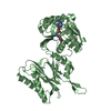
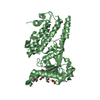
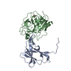
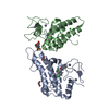
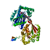
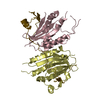
- PDB-3tm4: Crystal structure of Trm14 from Pyrococcus furiosus in complex wi... -
+ Open data

- Basic information
| Entry | Database: PDB / ID: 3tm4 | ||||||
|---|---|---|---|---|---|---|---|
| Title | Crystal structure of Trm14 from Pyrococcus furiosus in complex with S-adenosylmethionine | ||||||
 Components Components | tRNA (guanine N2-)-methyltransferase Trm14 | ||||||
 Keywords Keywords |  TRANSFERASE / Rossmann fold / methyltransferase / THUMP domain / tRNA methyltransferase TRANSFERASE / Rossmann fold / methyltransferase / THUMP domain / tRNA methyltransferase | ||||||
| Function / homology |  Function and homology information Function and homology informationtRNA (guanine(6)-N2)-methyltransferase activity / tRNA (guanine6-N2)-methyltransferase / tRNA modification / methylation / RNA binding / cytoplasmSimilarity search - Function | ||||||
| Biological species |   Pyrococcus furiosus (archaea) Pyrococcus furiosus (archaea) | ||||||
| Method | X-RAY DIFFRACTION / SYNCHROTRON / MOLECULAR REPLACEMENT / Resolution: 1.95 Å | ||||||
 Authors Authors | Fislage, M. / Roovers, M. / Tuszynska, I. / Bujnicki, J.M. / Droogmans, L. / Versees, W. | ||||||
 Citation Citation | Journal: Nucleic Acids Res. / Year: 2012 Title: Crystal structures of the tRNA:m2G6 methyltransferase Trm14/TrmN from two domains of life. Authors: Fislage, M. / Roovers, M. / Tuszynska, I. / Bujnicki, J.M. / Droogmans, L. / Versees, W. | ||||||
| History |
|
- Structure visualization
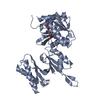
| Structure viewer | Molecule: MolmilJmol/JSmol |
|---|
- Downloads & links
-Download
| PDBx/mmCIF format | 3tm4.cif.gz | 166.1 KB | Display | PDBx/mmCIF format |
|---|---|---|---|---|
| PDB format | pdb3tm4.ent.gz | 131.2 KB | Display | PDB format |
| PDBx/mmJSON format | 3tm4.json.gz | Tree view | PDBx/mmJSON format | |
| Others |  Other downloads Other downloads |
-Validation report
| Arichive directory | https://data.pdbj.org/pub/pdb/validation_reports/tm/3tm4ftp://data.pdbj.org/pub/pdb/validation_reports/tm/3tm4 | HTTPS FTP |
|---|
-Related structure data
| Related structure data |  3tljSC  3tm5C  3tmaC S: Starting model for refinement C: citing same article ( |
|---|---|
| Similar structure data |


-Links
 PDBj
PDBj
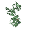
- Assembly
| Deposited unit | 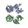
| ||||||||
|---|---|---|---|---|---|---|---|---|---|
| 1 | 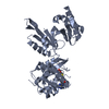
| ||||||||
| 2 | 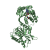
| ||||||||
| Unit cell |
| ||||||||
| Details | The asymmetric unit contains two identical molecules connected by NCS |
-Components
| #1: Protein | Mass: 42131.691 Da / Num. of mol.: 2 Source method: isolated from a genetically manipulated source Source: (gene. exp.) Pyrococcus furiosus (archaea) / Strain: ATCC 43587 / DSM 3638 / JCM 8422 / Vc1 / Gene: PF1002 / Plasmid: pET30 / Production host:  Escherichia coli (E. coli) / Strain (production host): Rosetta (DE3) Escherichia coli (E. coli) / Strain (production host): Rosetta (DE3)References: UniProt: Q8U248, Transferases; Transferring one-carbon groups; Methyltransferases#2: Chemical | S-Adenosyl methionine  Mass: 398.437 Da / Num. of mol.: 2 / Source method: obtained synthetically / Formula: C15H22N6O5S Mass: 398.437 Da / Num. of mol.: 2 / Source method: obtained synthetically / Formula: C15H22N6O5S#3: Water | ChemComp-HOH / | Water Mass: 18.015 Da / Num. of mol.: 382 / Source method: isolated from a natural source / Formula: H2O Mass: 18.015 Da / Num. of mol.: 382 / Source method: isolated from a natural source / Formula: H2O |
|---|
-Experimental details
-Experiment
| Experiment | Method: X-RAY DIFFRACTION / Number of used crystals: 1 |
|---|
- Sample preparation
| Crystal | Density Matthews: 2.67 Å3/Da / Density % sol: 53.93 % |
|---|---|
| Crystal grow | Temperature: 293 K / Method: vapor diffusion, hanging drop / pH: 8 Details: 100 mM Tris-acetate, 32% PEG 4000, 15% glycerol, crystal was soaked in crystallization solution containing 1 mM S-adenosylmethionine, pH 8, VAPOR DIFFUSION, HANGING DROP, temperature 293K |
-Data collection
| Diffraction | Mean temperature: 100 K | |||||||||||||||
|---|---|---|---|---|---|---|---|---|---|---|---|---|---|---|---|---|
| Diffraction source | Source: SYNCHROTRON / Site: SLS  / Beamline: X06DA / Wavelength: 1.0015 Å / Beamline: X06DA / Wavelength: 1.0015 Å | |||||||||||||||
| Detector | Type: MARMOSAIC 225 mm CCD / Detector: CCD / Date: Feb 25, 2011 | |||||||||||||||
| Radiation | Monochromator: Bartels Monochromator with dual channel cut crystals (DCCM) in (+--+) geometry Protocol: SINGLE WAVELENGTH / Monochromatic (M) / Laue (L): M / Scattering type: x-ray | |||||||||||||||
| Radiation wavelength | Wavelength: 1.0015 Å / Relative weight: 1 | |||||||||||||||
| Reflection twin |
| |||||||||||||||
| Reflection | Resolution: 1.95→50 Å / Num. all: 65622 / Num. obs: 65283 / % possible obs: 99.5 % / Observed criterion σ(F): 0 / Observed criterion σ(I): 0 / Redundancy: 7.1 % / Rmerge(I) obs: 0.073 / Net I/σ(I): 18.29 | |||||||||||||||
| Reflection shell | Resolution: 1.95→2 Å / Redundancy: 6.6 % / Rmerge(I) obs: 0.544 / Mean I/σ(I) obs: 3.6 / % possible all: 98.9 |
- Processing
| Software |
| ||||||||||||||||||||||||||||||||||||||||||||||||||||||||||||||||||||||||||||||||||||||||||||||||||||||||||||||||||||||||||||||||||||||||||||||||||||||||||||||||||||||||||
|---|---|---|---|---|---|---|---|---|---|---|---|---|---|---|---|---|---|---|---|---|---|---|---|---|---|---|---|---|---|---|---|---|---|---|---|---|---|---|---|---|---|---|---|---|---|---|---|---|---|---|---|---|---|---|---|---|---|---|---|---|---|---|---|---|---|---|---|---|---|---|---|---|---|---|---|---|---|---|---|---|---|---|---|---|---|---|---|---|---|---|---|---|---|---|---|---|---|---|---|---|---|---|---|---|---|---|---|---|---|---|---|---|---|---|---|---|---|---|---|---|---|---|---|---|---|---|---|---|---|---|---|---|---|---|---|---|---|---|---|---|---|---|---|---|---|---|---|---|---|---|---|---|---|---|---|---|---|---|---|---|---|---|---|---|---|---|---|---|---|---|---|
| Refinement | Method to determine structure: MOLECULAR REPLACEMENT Starting model: 3TLJ Resolution: 1.95→49.53 Å / Cor.coef. Fo:Fc: 0.962 / Cor.coef. Fo:Fc free: 0.946 / Isotropic thermal model: isotropic / Cross valid method: THROUGHOUT / ESU R: 0.031 / ESU R Free: 0.028 / Stereochemistry target values: MAXIMUM LIKELIHOOD / Details: HYDROGENS HAVE BEEN ADDED IN THE RIDING POSITIONS
| ||||||||||||||||||||||||||||||||||||||||||||||||||||||||||||||||||||||||||||||||||||||||||||||||||||||||||||||||||||||||||||||||||||||||||||||||||||||||||||||||||||||||||
| Solvent computation | Ion probe radii: 0.8 Å / Shrinkage radii: 0.8 Å / VDW probe radii: 1.4 Å / Solvent model: MASK | ||||||||||||||||||||||||||||||||||||||||||||||||||||||||||||||||||||||||||||||||||||||||||||||||||||||||||||||||||||||||||||||||||||||||||||||||||||||||||||||||||||||||||
| Displacement parameters | Biso mean: 29.15 Å2
| ||||||||||||||||||||||||||||||||||||||||||||||||||||||||||||||||||||||||||||||||||||||||||||||||||||||||||||||||||||||||||||||||||||||||||||||||||||||||||||||||||||||||||
| Refine analyze | Luzzati coordinate error obs: 0.2123 Å | ||||||||||||||||||||||||||||||||||||||||||||||||||||||||||||||||||||||||||||||||||||||||||||||||||||||||||||||||||||||||||||||||||||||||||||||||||||||||||||||||||||||||||
| Refinement step | Cycle: LAST / Resolution: 1.95→49.53 Å
| ||||||||||||||||||||||||||||||||||||||||||||||||||||||||||||||||||||||||||||||||||||||||||||||||||||||||||||||||||||||||||||||||||||||||||||||||||||||||||||||||||||||||||
| Refine LS restraints |
|
