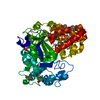
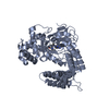
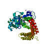
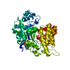
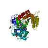
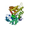
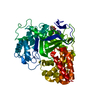
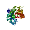
[English] 日本語
Yorodumi
- PDB-3tg4: Structure of SMYD2 in complex with SAM -

+
Open data


ID or keywords:

Loading...

-
Basic information

Entry
Database: PDB / ID: 3tg4
TitleStructure of SMYD2 in complex with SAM
ComponentsN-lysine methyltransferase SMYD2
KeywordsTRANSFERASE / SET domain / methyltransferase / co factor binding
Function / homology
Function and homology information


lysine N-methyltransferase activity / [histone H3]-lysine4 N-trimethyltransferase / peptidyl-lysine monomethylation / peptidyl-lysine dimethylation / histone H3K36 methyltransferase activity / histone H3K4 trimethyltransferase activity / protein-lysine N-methyltransferase activity / histone H3 methyltransferase activity / regulation of DNA damage response, signal transduction by p53 class mediator / RNA polymerase II complex binding ...lysine N-methyltransferase activity / [histone H3]-lysine4 N-trimethyltransferase / peptidyl-lysine monomethylation / peptidyl-lysine dimethylation / histone H3K36 methyltransferase activity / histone H3K4 trimethyltransferase activity / protein-lysine N-methyltransferase activity / histone H3 methyltransferase activity / regulation of DNA damage response, signal transduction by p53 class mediator / RNA polymerase II complex binding / Transferases; Transferring one-carbon groups; Methyltransferases / regulation of signal transduction by p53 class mediator / Regulation of TP53 Activity through Methylation / PKMTs methylate histone lysines / p53 binding / heart development / negative regulation of cell population proliferation / negative regulation of transcription by RNA polymerase II / nucleoplasm / metal ion binding / nucleus / cytosol / cytoplasm
Similarity search - Function
SMYD2, SET domain / MYND finger / Zinc finger, MYND-type / Zinc finger MYND-type signature. / Zinc finger MYND-type profile. / Beta-clip-like / SET domain / Tetratricopeptide repeat domain / SET (Su(var)3-9, Enhancer-of-zeste, Trithorax) domain / SET domain superfamily ...SMYD2, SET domain / MYND finger / Zinc finger, MYND-type / Zinc finger MYND-type signature. / Zinc finger MYND-type profile. / Beta-clip-like / SET domain / Tetratricopeptide repeat domain / SET (Su(var)3-9, Enhancer-of-zeste, Trithorax) domain / SET domain superfamily / SET domain / SET domain profile. / SET domain / Beta Complex / Serine Threonine Protein Phosphatase 5, Tetratricopeptide repeat / Alpha Horseshoe / Tetratricopeptide-like helical domain superfamily / Mainly Beta / Mainly Alpha
Similarity search - Domain/homology
S-ADENOSYLMETHIONINE / N-lysine methyltransferase SMYD2
Similarity search - Component
Biological speciesHomo sapiens (human)
MethodX-RAY DIFFRACTION / SYNCHROTRON / MOLECULAR REPLACEMENT / Resolution: 2 Å
AuthorsZhao, K. / Wang, L.
CitationJournal: J.Biol.Chem. / Year: 2011
Title: Structure of human SMYD2 reveals the basis of p53 tumor suppressor methylation
Authors: Wang, L. / Li, L. / Zhang, H. / Luo, X. / Dai, J. / Zhou, S. / Gu, J. / Zhu, J. / Atadja, P. / Lu, C. / Li, E. / Zhao, K.
History
DepositionAug 17, 2011Deposition site: RCSB / Processing site: PDBJ
Revision 1.0Aug 31, 2011Provider: repository / Type: Initial release
Revision 1.1Sep 14, 2011Group: Database references
Revision 1.2Nov 1, 2023Group: Data collection / Database references ...Data collection / Database references / Derived calculations / Refinement description
Category: chem_comp_atom / chem_comp_bond ...chem_comp_atom / chem_comp_bond / database_2 / pdbx_initial_refinement_model / pdbx_struct_conn_angle / struct_conn / struct_site
Item: _database_2.pdbx_DOI / _database_2.pdbx_database_accession ..._database_2.pdbx_DOI / _database_2.pdbx_database_accession / _pdbx_struct_conn_angle.ptnr1_auth_comp_id / _pdbx_struct_conn_angle.ptnr1_auth_seq_id / _pdbx_struct_conn_angle.ptnr1_label_atom_id / _pdbx_struct_conn_angle.ptnr1_label_comp_id / _pdbx_struct_conn_angle.ptnr1_label_seq_id / _pdbx_struct_conn_angle.ptnr2_auth_seq_id / _pdbx_struct_conn_angle.ptnr2_label_asym_id / _pdbx_struct_conn_angle.ptnr3_auth_comp_id / _pdbx_struct_conn_angle.ptnr3_auth_seq_id / _pdbx_struct_conn_angle.ptnr3_label_atom_id / _pdbx_struct_conn_angle.ptnr3_label_comp_id / _pdbx_struct_conn_angle.ptnr3_label_seq_id / _pdbx_struct_conn_angle.value / _struct_conn.pdbx_dist_value / _struct_conn.ptnr1_auth_comp_id / _struct_conn.ptnr1_auth_seq_id / _struct_conn.ptnr1_label_atom_id / _struct_conn.ptnr1_label_comp_id / _struct_conn.ptnr1_label_seq_id / _struct_conn.ptnr2_auth_seq_id / _struct_conn.ptnr2_label_asym_id / _struct_site.pdbx_auth_asym_id / _struct_site.pdbx_auth_comp_id / _struct_site.pdbx_auth_seq_id

-
Structure visualization

Structure viewerMolecule:
MolmilJmol/JSmol

Downloads & links

-
Assembly

Deposited unit
A: N-lysine methyltransferase SMYD2
hetero molecules


Theoretical massNumber of molelcules
Total (without water)50,81510
Polymers49,7601
Non-polymers1,0559
Water4,270237
1


  • Idetical with deposited unit
  • defined by author&software
TypeNameSymmetry operationNumber
identity operation1_555x,y,z1
MethodPISA
Unit cell
Length a, b, c (Å)52.480, 67.480, 141.970
Angle α, β, γ (deg.)90.00, 90.00, 90.00
Int Tables number19
Space group name H-MP212121

-
Components

#1: Protein N-lysine methyltransferase SMYD2 / HSKM-B / Histone methyltransferase SMYD2 / Lysine N-methyltransferase 3C / SET and MYND domain- ...HSKM-B / Histone methyltransferase SMYD2 / Lysine N-methyltransferase 3C / SET and MYND domain-containing protein 2


Mass: 49760.102 Da / Num. of mol.: 1
Source method: isolated from a genetically manipulated source
Source: (gene. exp.) Homo sapiens (human) / Gene: SMYD2, KMT3C / Plasmid: pFastbac / Cell line (production host): SF9 / Production host: Spodoptera frugiperda (fall armyworm)
References: UniProt: Q9NRG4, Transferases; Transferring one-carbon groups; Methyltransferases, histone-lysine N-methyltransferase
#2: Chemical ChemComp-SAM / S-ADENOSYLMETHIONINE / S-Adenosyl methionine


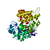
Mass: 398.437 Da / Num. of mol.: 1 / Source method: obtained synthetically / Formula: C15H22N6O5S
#3: Chemical ChemComp-ZN / ZINC ION


Mass: 65.409 Da / Num. of mol.: 3 / Source method: obtained synthetically / Formula: Zn
#4: Chemical
ChemComp-GOL / GLYCEROL / GLYCERIN / PROPANE-1,2,3-TRIOL / Glycerol


Mass: 92.094 Da / Num. of mol.: 5 / Source method: obtained synthetically / Formula: C3H8O3
#5: Water ChemComp-HOH / water / Water


Mass: 18.015 Da / Num. of mol.: 237 / Source method: isolated from a natural source / Formula: H2O

-
Experimental details

-
Experiment

ExperimentMethod: X-RAY DIFFRACTION / Number of used crystals: 1

-
Sample preparation

CrystalDensity Matthews: 2.53 Å3/Da / Density % sol: 51.31 %
Crystal growTemperature: 293 K / Method: vapor diffusion / pH: 8.5
Details: 0.8M lithium chloride, 0.1M Tris-HCl (pH 8.5), 32% PEG 4000, VAPOR DIFFUSION, temperature 293K

-
Data collection

DiffractionMean temperature: 100 K
Diffraction sourceSource: SYNCHROTRON / Site: SSRF / Beamline: BL17U / Wavelength: 0.97924 Å
DetectorType: ADSC QUANTUM 4r / Detector: CCD / Date: May 10, 2011
RadiationMonochromator: Toroidal mirror / Protocol: SINGLE WAVELENGTH / Monochromatic (M) / Laue (L): M / Scattering type: x-ray
Radiation wavelengthWavelength: 0.97924 Å / Relative weight: 1
ReflectionResolution: 2→70.99 Å / Num. all: 34948 / Num. obs: 34884 / % possible obs: 100 % / Observed criterion σ(F): 2 / Observed criterion σ(I): 2
Reflection shellResolution: 2→2.11 Å / % possible all: 100

-
Processing

Software
NameVersionClassification
HKL-2000data collection
MOLREPphasing
REFMAC5.5.0109refinement
HKL-2000data reduction
SCALAdata scaling
RefinementMethod to determine structure: MOLECULAR REPLACEMENT
Starting model: PDB ENTRY 3MEK
Resolution: 2→19.88 Å / Cor.coef. Fo:Fc: 0.955 / Cor.coef. Fo:Fc free: 0.936 / SU B: 8.138 / SU ML: 0.116 / Cross valid method: THROUGHOUT / ESU R Free: 0.154 / Stereochemistry target values: MAXIMUM LIKELIHOOD / Details: HYDROGENS HAVE BEEN ADDED IN THE RIDING POSITIONS
RfactorNum. reflection% reflectionSelection details
Rfree0.2244 1749 5 %RANDOM
Rwork0.18966 ---
all0.19142 34753 --
obs0.19142 33004 99.73 %-
Solvent computationIon probe radii: 0.8 Å / Shrinkage radii: 0.8 Å / VDW probe radii: 1.4 Å / Solvent model: MASK
Displacement parametersBiso mean: 43.464 Å2
Baniso -1Baniso -2Baniso -3
1--0.14 Å20 Å20 Å2
2--0.17 Å20 Å2
3----0.04 Å2
Refinement stepCycle: LAST / Resolution: 2→19.88 Å
ProteinNucleic acidLigandSolventTotal
Num. atoms3434 0 60 237 3731
Refine LS restraints
Refine-IDTypeDev idealDev ideal targetNumber
X-RAY DIFFRACTIONr_bond_refined_d0.010.0223578
X-RAY DIFFRACTIONr_angle_refined_deg1.1791.9824812
X-RAY DIFFRACTIONr_dihedral_angle_1_deg5.5165426
X-RAY DIFFRACTIONr_dihedral_angle_2_deg35.03224.251167
X-RAY DIFFRACTIONr_dihedral_angle_3_deg15.23215645
X-RAY DIFFRACTIONr_dihedral_angle_4_deg15.7151520
X-RAY DIFFRACTIONr_chiral_restr0.0790.2509
X-RAY DIFFRACTIONr_gen_planes_refined0.0050.0212668
X-RAY DIFFRACTIONr_mcbond_it0.4471.52136
X-RAY DIFFRACTIONr_mcangle_it0.86723441
X-RAY DIFFRACTIONr_scbond_it1.77731442
X-RAY DIFFRACTIONr_scangle_it2.6754.51371
LS refinement shellResolution: 2→2.051 Å / Total num. of bins used: 20
RfactorNum. reflection% reflection
Rfree0.279 120 -
Rwork0.228 2381 -
obs--99.56 %
Refinement TLS params.

Method: refined / Refine-ID: X-RAY DIFFRACTION

IDL112)L122)L132)L222)L232)L332)S11 (Å °)S12 (Å °)S13 (Å °)S21 (Å °)S22 (Å °)S23 (Å °)S31 (Å °)S32 (Å °)S33 (Å °)T112)T122)T132)T222)T232)T332)Origin x (Å)Origin y (Å)Origin z (Å)
11.3301-0.6188-0.76724.16791.63022.21030.15680.02040.0369-0.29950.1383-0.469-0.31260.0689-0.2950.0329-0.01190.10440.0364-0.03450.07311.2456.02820.434
210.92112.0211-6.79754.31710.523710.07990.00710.7570.5508-0.90040.06880.5098-1.2836-0.4977-0.07590.99730.3909-0.21070.37770.04650.1802-2.8141.233-7.245
32.9501-0.12920.80996.23090.89332.90680.2710.0956-0.1709-0.5755-0.29470.29480.1271-0.20060.02370.08580.05560.00720.1087-0.09530.0752.57-14.3839.344
Refinement TLS group
IDRefine-IDRefine TLS-IDAuth asym-IDAuth seq-ID
1X-RAY DIFFRACTION1A6 - 276
2X-RAY DIFFRACTION2A277 - 334
3X-RAY DIFFRACTION3A335 - 432

+
About Yorodumi

-
News

-
Feb 9, 2022. New format data for meta-information of EMDB entries

New format data for meta-information of EMDB entries

  • Version 3 of the EMDB header file is now the official format.
  • The previous official version 1.9 will be removed from the archive.

Related info.:EMDB header

External links:wwPDB to switch to version 3 of the EMDB data model

-
Aug 12, 2020. Covid-19 info

Covid-19 info

URL: https://pdbj.org/emnavi/covid19.php

New page: Covid-19 featured information page in EM Navigator.

Related info.:Covid-19 info / Mar 5, 2020. Novel coronavirus structure data

+
Mar 5, 2020. Novel coronavirus structure data

Novel coronavirus structure data

Related info.:Yorodumi Speices / Aug 12, 2020. Covid-19 info

External links:COVID-19 featured content - PDBj / Molecule of the Month (242):Coronavirus Proteases

+
Jan 31, 2019. EMDB accession codes are about to change! (news from PDBe EMDB page)

EMDB accession codes are about to change! (news from PDBe EMDB page)

  • The allocation of 4 digits for EMDB accession codes will soon come to an end. Whilst these codes will remain in use, new EMDB accession codes will include an additional digit and will expand incrementally as the available range of codes is exhausted. The current 4-digit format prefixed with “EMD-” (i.e. EMD-XXXX) will advance to a 5-digit format (i.e. EMD-XXXXX), and so on. It is currently estimated that the 4-digit codes will be depleted around Spring 2019, at which point the 5-digit format will come into force.
  • The EM Navigator/Yorodumi systems omit the EMD- prefix.

Related info.:Q: What is EMD? / ID/Accession-code notation in Yorodumi/EM Navigator

External links:EMDB Accession Codes are Changing Soon! / Contact to PDBj

+
Jul 12, 2017. Major update of PDB

Major update of PDB

  • wwPDB released updated PDB data conforming to the new PDBx/mmCIF dictionary.
  • This is a major update changing the version number from 4 to 5, and with Remediation, in which all the entries are updated.
  • In this update, many items about electron microscopy experimental information are reorganized (e.g. em_software).
  • Now, EM Navigator and Yorodumi are based on the updated data.

External links:wwPDB Remediation / Enriched Model Files Conforming to OneDep Data Standards Now Available in the PDB FTP Archive

-
Yorodumi

Thousand views of thousand structures

  • Yorodumi is a browser for structure data from EMDB, PDB, SASBDB, etc.
  • This page is also the successor to EM Navigator detail page, and also detail information page/front-end page for Omokage search.
  • The word "yorodu" (or yorozu) is an old Japanese word meaning "ten thousand". "mi" (miru) is to see.

Related info.:EMDB / PDB / SASBDB / Comparison of 3 databanks / Yorodumi Search / Aug 31, 2016. New EM Navigator & Yorodumi / Yorodumi Papers / Jmol/JSmol / Function and homology information / Changes in new EM Navigator and Yorodumi

Read more