 Movie
Movie Controller

[English] 日本語
 Yorodumi
- PDB-3tdn: Computationally designed two-fold symmetric Tim-barrel protein, FLR -
+ Open data

- Basic information
| Entry | Database: PDB / ID: 3tdn | ||||||
|---|---|---|---|---|---|---|---|
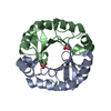
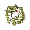
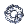
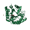
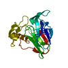
| Title | Computationally designed two-fold symmetric Tim-barrel protein, FLR | ||||||
 Components Components | FLR SYMMETRIC ALPHA-BETA TIM BARREL | ||||||
 Keywords Keywords |  DE NOVO PROTEIN / SYMMETRIC SUPERFOLD / TIM-BARREL DE NOVO PROTEIN / SYMMETRIC SUPERFOLD / TIM-BARREL | ||||||
| Function / homology | Rossmann fold - #12600 / Aldolase class I / TIM Barrel / Alpha-Beta Barrel / Rossmann fold / 3-Layer(aba) Sandwich / Alpha Beta Function and homology information Function and homology information | ||||||
| Biological species | synthetic construct (others) | ||||||
| Method | X-RAY DIFFRACTION / SYNCHROTRON / MOLECULAR REPLACEMENT / Resolution: 1.4 Å | ||||||
 Authors Authors | Harp, J.M. / Fortenberry, C. / Bowman, E. / Profitt, W. / Dorr, B. / Mizoue, L. / Meiler, J. | ||||||
 Citation Citation | Journal: J.Am.Chem.Soc. / Year: 2011 Title: Exploring symmetry as an avenue to the computational design of large protein domains. Authors: Fortenberry, C. / Bowman, E.A. / Proffitt, W. / Dorr, B. / Combs, S. / Harp, J. / Mizoue, L. / Meiler, J. | ||||||
| History |
|
- Structure visualization
| Structure viewer | Molecule: MolmilJmol/JSmol |
|---|
- Downloads & links
-Download
| PDBx/mmCIF format | 3tdn.cif.gz | 158.5 KB | Display | PDBx/mmCIF format |
|---|---|---|---|---|
| PDB format | pdb3tdn.ent.gz | 126 KB | Display | PDB format |
| PDBx/mmJSON format | 3tdn.json.gz | Tree view | PDBx/mmJSON format | |
| Others |  Other downloads Other downloads |
-Validation report
| Arichive directory | https://data.pdbj.org/pub/pdb/validation_reports/td/3tdnftp://data.pdbj.org/pub/pdb/validation_reports/td/3tdn | HTTPS FTP |
|---|
-Related structure data


-Links
 PDBj
PDBj
- Assembly
| Deposited unit | 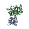
| ||||||||
|---|---|---|---|---|---|---|---|---|---|
| 1 | 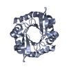
| ||||||||
| 2 | 
| ||||||||
| Unit cell |
|
-Components
| #1: Protein | Mass: 26697.471 Da / Num. of mol.: 2 Source method: isolated from a genetically manipulated source Source: (gene. exp.) synthetic construct (others) / Production host:  ESCHERICHIA COLI (E. coli) ESCHERICHIA COLI (E. coli)#2: Water | ChemComp-HOH / | Water Mass: 18.015 Da / Num. of mol.: 259 / Source method: isolated from a natural source / Formula: H2O Mass: 18.015 Da / Num. of mol.: 259 / Source method: isolated from a natural source / Formula: H2OSequence details | THE SEQUENCE IS COMPUTATIONALLY DESIGNED BASED ON A TIM-BARREL. THE FULL CRYSTALLIZED SEQUENCE HAS ...THE SEQUENCE IS COMPUTATIO | |
|---|
-Experimental details
-Experiment
| Experiment | Method: X-RAY DIFFRACTION / Number of used crystals: 1 |
|---|
- Sample preparation
| Crystal | Density Matthews: 2.07 Å3/Da / Density % sol: 40.7 % |
|---|---|
| Crystal grow | Temperature: 291 K / Method: microbatch / pH: 4.2 Details: 20% PEG 1000, 0.1M SODIUM CITRATE, 0.1 M AMMONIUM CHLORIDE, pH 4.2, Microbatch, temperature 291K |
-Data collection
| Diffraction | Mean temperature: 100 K |
|---|---|
| Diffraction source | Source: SYNCHROTRON / Site: ALS  / Beamline: 12.3.1 / Wavelength: 1.072 / Beamline: 12.3.1 / Wavelength: 1.072 |
| Detector | Type: ADSC QUANTUM 315 / Detector: CCD / Date: Nov 23, 2009 |
| Radiation | Monochromator: KOHZU DUAL DOUBLE CRYSTAL MONOCHROMATOR / Protocol: SINGLE WAVELENGTH / Monochromatic (M) / Laue (L): M / Scattering type: x-ray |
| Radiation wavelength | Wavelength: 1.072 Å / Relative weight: 1 |
| Reflection | Resolution: 1.4→50 Å / Num. obs: 63971 / % possible obs: 98 % / Redundancy: 9.7 % / Biso Wilson estimate: 21 Å2 / Rmerge(I) obs: 0.072 / Net I/σ(I): 19.35 |
| Reflection shell | Resolution: 1.4→1.45 Å / Redundancy: 9.6 % / Rmerge(I) obs: 0.683 / Mean I/σ(I) obs: 3.01 / % possible all: 96.9 |
- Processing
| Software |
| ||||||||||||||||||||||||||||||||||||||||||||||||||||||||||||||||||||||||||||||||||||||||||||||||||||||||||||||||||||||||||||||||||||||||||||||||||||||||||||||||||||||||||
|---|---|---|---|---|---|---|---|---|---|---|---|---|---|---|---|---|---|---|---|---|---|---|---|---|---|---|---|---|---|---|---|---|---|---|---|---|---|---|---|---|---|---|---|---|---|---|---|---|---|---|---|---|---|---|---|---|---|---|---|---|---|---|---|---|---|---|---|---|---|---|---|---|---|---|---|---|---|---|---|---|---|---|---|---|---|---|---|---|---|---|---|---|---|---|---|---|---|---|---|---|---|---|---|---|---|---|---|---|---|---|---|---|---|---|---|---|---|---|---|---|---|---|---|---|---|---|---|---|---|---|---|---|---|---|---|---|---|---|---|---|---|---|---|---|---|---|---|---|---|---|---|---|---|---|---|---|---|---|---|---|---|---|---|---|---|---|---|---|---|---|---|
| Refinement | Method to determine structure: MOLECULAR REPLACEMENT Starting model: COMPUTATIONAL MODEL Resolution: 1.4→42.5 Å / Cor.coef. Fo:Fc: 0.968 / Cor.coef. Fo:Fc free: 0.958 / SU B: 1.793 / SU ML: 0.028 / Cross valid method: THROUGHOUT / ESU R Free: 0.013 / Stereochemistry target values: MAXIMUM LIKELIHOOD / Details: HYDROGENS HAVE BEEN ADDED IN THE RIDING POSITIONS
| ||||||||||||||||||||||||||||||||||||||||||||||||||||||||||||||||||||||||||||||||||||||||||||||||||||||||||||||||||||||||||||||||||||||||||||||||||||||||||||||||||||||||||
| Solvent computation | Solvent model: BABINET MODEL PARAMETERS FOR MASK CACLULATION | ||||||||||||||||||||||||||||||||||||||||||||||||||||||||||||||||||||||||||||||||||||||||||||||||||||||||||||||||||||||||||||||||||||||||||||||||||||||||||||||||||||||||||
| Displacement parameters | Biso mean: 21.27 Å2
| ||||||||||||||||||||||||||||||||||||||||||||||||||||||||||||||||||||||||||||||||||||||||||||||||||||||||||||||||||||||||||||||||||||||||||||||||||||||||||||||||||||||||||
| Refinement step | Cycle: LAST / Resolution: 1.4→42.5 Å
| ||||||||||||||||||||||||||||||||||||||||||||||||||||||||||||||||||||||||||||||||||||||||||||||||||||||||||||||||||||||||||||||||||||||||||||||||||||||||||||||||||||||||||
| Refine LS restraints |
| ||||||||||||||||||||||||||||||||||||||||||||||||||||||||||||||||||||||||||||||||||||||||||||||||||||||||||||||||||||||||||||||||||||||||||||||||||||||||||||||||||||||||||
| LS refinement shell | Resolution: 1.4→1.44 Å / Total num. of bins used: 20
|
