 Movie
Movie Controller

[English] 日本語
 Yorodumi
- PDB-3sf4: Crystal structure of the complex between the conserved cell polar... -
+ Open data

- Basic information
| Entry | Database: PDB / ID: 3sf4 | ||||||
|---|---|---|---|---|---|---|---|
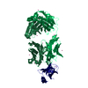
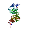
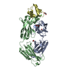
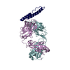
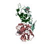
| Title | Crystal structure of the complex between the conserved cell polarity proteins Inscuteable and LGN | ||||||
 Components Components |
| ||||||
 Keywords Keywords |  SIGNALING PROTEIN/PROTEIN BINDING / tetratricopeptide repeat / TPR / cell polarity / asymmetric cell division / mitotic spindle orientation / Cytoplasm and cell cortex / SIGNALING PROTEIN-PROTEIN BINDING complex SIGNALING PROTEIN/PROTEIN BINDING / tetratricopeptide repeat / TPR / cell polarity / asymmetric cell division / mitotic spindle orientation / Cytoplasm and cell cortex / SIGNALING PROTEIN-PROTEIN BINDING complex | ||||||
| Function / homology |  Function and homology information Function and homology informationlateral cell cortex / cell cortex region / maintenance of centrosome location / asymmetric cell division / positive regulation of spindle assembly / GDP-dissociation inhibitor activity / dynein complex binding / mitotic spindle pole / establishment of mitotic spindle orientation / positive regulation of protein localization to cell cortex ...lateral cell cortex / cell cortex region / maintenance of centrosome location / asymmetric cell division / positive regulation of spindle assembly / GDP-dissociation inhibitor activity / dynein complex binding / mitotic spindle pole / establishment of mitotic spindle orientation / positive regulation of protein localization to cell cortex / G-protein alpha-subunit binding / lateral plasma membrane / regulation of mitotic spindle organization / mitotic spindle organization / regulation of protein stability / : / protein-macromolecule adaptor activity / nervous system development / cell cortex / G alpha (i) signalling events / cell differentiation / G protein-coupled receptor signaling pathway / cell division / protein domain specific binding / nucleotide binding / centrosome / protein-containing complex / identical protein binding / plasma membrane / cytosol / cytoplasmSimilarity search - Function | ||||||
| Biological species |  Homo sapiens (human) Homo sapiens (human) | ||||||
| Method | X-RAY DIFFRACTION / SYNCHROTRON / MOLECULAR REPLACEMENT / Resolution: 2.6 Å | ||||||
 Authors Authors | Yuzawa, S. / Kamakura, S. / Iwakiri, Y. / Hayase, J. / Sumimoto, H. | ||||||
 Citation Citation | Journal: Proc.Natl.Acad.Sci.USA / Year: 2011 Title: Structural basis for interaction between the conserved cell polarity proteins Inscuteable and Leu-Gly-Asn repeat-enriched protein (LGN) Authors: Yuzawa, S. / Kamakura, S. / Iwakiri, Y. / Hayase, J. / Sumimoto, H. | ||||||
| History |
|
- Structure visualization
| Structure viewer | Molecule: MolmilJmol/JSmol |
|---|
- Downloads & links
-Download
| PDBx/mmCIF format | 3sf4.cif.gz | 236.3 KB | Display | PDBx/mmCIF format |
|---|---|---|---|---|
| PDB format | pdb3sf4.ent.gz | 189.2 KB | Display | PDB format |
| PDBx/mmJSON format | 3sf4.json.gz | Tree view | PDBx/mmJSON format | |
| Others |  Other downloads Other downloads |
-Validation report
| Arichive directory | https://data.pdbj.org/pub/pdb/validation_reports/sf/3sf4ftp://data.pdbj.org/pub/pdb/validation_reports/sf/3sf4 | HTTPS FTP |
|---|
-Related structure data


-Links
 PDBj
PDBj


- Assembly
| Deposited unit | 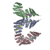
| ||||||||
|---|---|---|---|---|---|---|---|---|---|
| 1 | 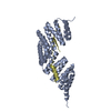
| ||||||||
| 2 | 
| ||||||||
| 3 | 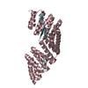
| ||||||||
| Unit cell |
|
-Components
| #1: Protein | Mass: 44948.281 Da / Num. of mol.: 3 / Fragment: N-terminal TPR domain (residues in UNP 20-421) Source method: isolated from a genetically manipulated source Source: (gene. exp.) Homo sapiens (human) / Gene: LGN / Plasmid: pRSFDuet-1 / Production host:  Escherichia coli (E. coli) / Strain (production host): BL21(DE3) / References: UniProt: P81274 Escherichia coli (E. coli) / Strain (production host): BL21(DE3) / References: UniProt: P81274#2: Protein | Mass: 5597.461 Da / Num. of mol.: 3 / Fragment: LGN-binding domain (residues in UNP 70-116) Source method: isolated from a genetically manipulated source Source: (gene. exp.) Homo sapiens (human) / Gene: INSC / Plasmid: pGEX-6p / Production host: Escherichia coli (E. coli) / Strain (production host): BL21(DE3) / References: UniProt: Q1MX18#3: Water | ChemComp-HOH / | Water Mass: 18.015 Da / Num. of mol.: 208 / Source method: isolated from a natural source / Formula: H2O Mass: 18.015 Da / Num. of mol.: 208 / Source method: isolated from a natural source / Formula: H2O |
|---|
-Experimental details
-Experiment
| Experiment | Method: X-RAY DIFFRACTION / Number of used crystals: 1 |
|---|
- Sample preparation
| Crystal | Density Matthews: 3 Å3/Da / Density % sol: 59.04 % |
|---|---|
| Crystal grow | Temperature: 293 K / Method: vapor diffusion, hanging drop / pH: 8.5 Details: 0.1M Tris HCl, 15% PEG 3350, 0.2M MgCL2, 4% 2,2,2-Trifluoroethanol, pH 8.5, VAPOR DIFFUSION, HANGING DROP, temperature 293K |
-Data collection
| Diffraction | Mean temperature: 95 K |
|---|---|
| Diffraction source | Source: SYNCHROTRON / Site: Photon Factory  / Beamline: BL-17A / Wavelength: 0.98 Å / Beamline: BL-17A / Wavelength: 0.98 Å |
| Detector | Type: ADSC QUANTUM 315r / Detector: CCD / Date: Jun 8, 2010 |
| Radiation | Protocol: SINGLE WAVELENGTH / Monochromatic (M) / Laue (L): M / Scattering type: x-ray |
| Radiation wavelength | Wavelength: 0.98 Å / Relative weight: 1 |
| Reflection | Resolution: 2.6→50 Å / Num. all: 57538 / Num. obs: 57538 / % possible obs: 100 % / Redundancy: 12.1 % / Biso Wilson estimate: 46.8 Å2 / Rmerge(I) obs: 0.096 |
| Reflection shell | Resolution: 2.6→2.64 Å / Redundancy: 10.4 % / Rmerge(I) obs: 0.422 / Mean I/σ(I) obs: 6.17 / Num. unique all: 2791 / % possible all: 100 |
- Processing
| Software |
| ||||||||||||||||||||||||||||||||||||
|---|---|---|---|---|---|---|---|---|---|---|---|---|---|---|---|---|---|---|---|---|---|---|---|---|---|---|---|---|---|---|---|---|---|---|---|---|---|
| Refinement | Method to determine structure: MOLECULAR REPLACEMENT Starting model: PDB ENTRY 2FO7, 2WQH, 1NA0 Resolution: 2.6→48.72 Å / Rfactor Rfree error: 0.005 / Data cutoff high absF: 3108570.8 / Data cutoff low absF: 0 / Isotropic thermal model: RESTRAINED / Cross valid method: THROUGHOUT / σ(F): 0 / Stereochemistry target values: Engh & Huber / Details: BULK SOLVENT MODEL USED
| ||||||||||||||||||||||||||||||||||||
| Solvent computation | Solvent model: FLAT MODEL / Bsol: 35.5476 Å2 / ksol: 0.35 e/Å3 | ||||||||||||||||||||||||||||||||||||
| Displacement parameters | Biso mean: 47.5 Å2
| ||||||||||||||||||||||||||||||||||||
| Refine analyze |
| ||||||||||||||||||||||||||||||||||||
| Refinement step | Cycle: LAST / Resolution: 2.6→48.72 Å
| ||||||||||||||||||||||||||||||||||||
| Refine LS restraints |
| ||||||||||||||||||||||||||||||||||||
| LS refinement shell | Resolution: 2.6→2.76 Å / Rfactor Rfree error: 0.014 / Total num. of bins used: 6
| ||||||||||||||||||||||||||||||||||||
| Xplor file |
|
