 Movie
Movie Controller


+ Open data

- Basic information
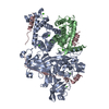
| Entry | Database: PDB / ID: 3sek | ||||||
|---|---|---|---|---|---|---|---|
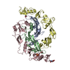
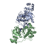
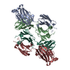
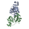
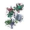
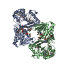
| Title | Crystal Structure of the Myostatin:Follistatin-like 3 Complex | ||||||
 Components Components |
| ||||||
 Keywords Keywords |  SIGNALING PROTEIN / protein-protein complex / TB domain / cystine knot motif / TGF-beta fold / disulfide linked dimer / follistatin domain (FSD) SIGNALING PROTEIN / protein-protein complex / TB domain / cystine knot motif / TGF-beta fold / disulfide linked dimer / follistatin domain (FSD) | ||||||
| Function / homology |  Function and homology information Function and homology informationnegative regulation of transmembrane receptor protein serine/threonine kinase signaling pathway / negative regulation of muscle hypertrophy / negative regulation of skeletal muscle tissue growth / negative regulation of myoblast proliferation / skeletal muscle satellite cell differentiation / myoblast migration involved in skeletal muscle regeneration / negative regulation of skeletal muscle satellite cell proliferation / negative regulation of satellite cell differentiation / Antagonism of Activin by Follistatin / skeletal muscle atrophy ...negative regulation of transmembrane receptor protein serine/threonine kinase signaling pathway / negative regulation of muscle hypertrophy / negative regulation of skeletal muscle tissue growth / negative regulation of myoblast proliferation / skeletal muscle satellite cell differentiation / myoblast migration involved in skeletal muscle regeneration / negative regulation of skeletal muscle satellite cell proliferation / negative regulation of satellite cell differentiation / Antagonism of Activin by Follistatin / skeletal muscle atrophy / ovulation cycle process / regulation of BMP signaling pathway / response to gravity / positive regulation of cell-cell adhesion / skeletal muscle tissue regeneration / activin binding / negative regulation of activin receptor signaling pathway / negative regulation of myoblast differentiation / response to muscle activity / muscle cell cellular homeostasis / negative regulation of kinase activity / positive regulation of macrophage chemotaxis / response to testosterone / negative regulation of osteoclast differentiation / fibronectin binding / negative regulation of BMP signaling pathway / negative regulation of phosphatidylinositol 3-kinase/protein kinase B signal transduction / hematopoietic progenitor cell differentiation / positive regulation of lamellipodium assembly / response to electrical stimulus / negative regulation of insulin receptor signaling pathway / cellular response to dexamethasone stimulus / ossification / transforming growth factor beta receptor signaling pathway / cytokine activity / Post-translational protein phosphorylation / growth factor activity / response to estrogen / Regulation of Insulin-like Growth Factor (IGF) transport and uptake by Insulin-like Growth Factor Binding Proteins (IGFBPs) / heparin binding / cellular response to hypoxia / response to ethanol / cell differentiation / endoplasmic reticulum lumen / signaling receptor binding / regulation of transcription by RNA polymerase II / positive regulation of DNA-templated transcription / protein homodimerization activity / positive regulation of transcription by RNA polymerase II / extracellular space / extracellular region / nucleoplasm / identical protein binding / nucleusSimilarity search - Function | ||||||
| Biological species |  Mus musculus (house mouse) Mus musculus (house mouse) Homo sapiens (human) Homo sapiens (human) | ||||||
| Method | X-RAY DIFFRACTION / SYNCHROTRON / MOLECULAR REPLACEMENT / molecular replacement / Resolution: 2.401 Å | ||||||
 Authors Authors | Cash, J.N. / Thompson, T.B. | ||||||
 Citation Citation | Journal: J.Biol.Chem. / Year: 2012 Title: Structure of myostatinfollistatin-like 3: N-terminal domains of follistatin-type molecules exhibit alternate modes of binding. Authors: Cash, J.N. / Angerman, E.B. / Kattamuri, C. / Nolan, K. / Zhao, H. / Sidis, Y. / Keutmann, H.T. / Thompson, T.B. | ||||||
| History |
|
- Structure visualization
| Structure viewer | Molecule: MolmilJmol/JSmol |
|---|
- Downloads & links
-Download
| PDBx/mmCIF format | 3sek.cif.gz | 138.2 KB | Display | PDBx/mmCIF format |
|---|---|---|---|---|
| PDB format | pdb3sek.ent.gz | 107.8 KB | Display | PDB format |
| PDBx/mmJSON format | 3sek.json.gz | Tree view | PDBx/mmJSON format | |
| Others |  Other downloads Other downloads |
-Validation report
| Arichive directory | https://data.pdbj.org/pub/pdb/validation_reports/se/3sekftp://data.pdbj.org/pub/pdb/validation_reports/se/3sek | HTTPS FTP |
|---|
-Related structure data


-Links
 PDBj
PDBj


- Assembly
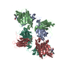
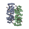
| Deposited unit | 
| ||||||||
|---|---|---|---|---|---|---|---|---|---|
| 1 | 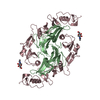
| ||||||||
| Unit cell |
|
-Components
| #1: Protein | Mass: 12422.212 Da / Num. of mol.: 1 / Fragment: UNP Residues 268-376 Source method: isolated from a genetically manipulated source Source: (gene. exp.) Mus musculus (house mouse) / Gene: Gdf8, Mstn, myostatin / Production host:  Cricetulus griseus (Chinese hamster) / References: UniProt: O08689 Cricetulus griseus (Chinese hamster) / References: UniProt: O08689 |
|---|---|
| #2: Protein | Mass: 22233.553 Da / Num. of mol.: 1 / Fragment: UNP Residues 36-244 Source method: isolated from a genetically manipulated source Source: (gene. exp.) Homo sapiens (human) / Gene: FLRG, follistatin-like 3, FSTL3, UNQ674/PRO1308 / Plasmid: pcDNA3.1 / Cell line (production host): HEK293 / Production host: Homo sapiens (human) / References: UniProt: O95633 |
| #3: Sugar | ChemComp-NAG / N-Acetylglucosamine  Type: D-saccharide, beta linking / Mass: 221.208 Da / Num. of mol.: 1 Type: D-saccharide, beta linking / Mass: 221.208 Da / Num. of mol.: 1Source method: isolated from a genetically manipulated source Formula: C8H15NO6 |
| #4: Water | ChemComp-HOH / Water Mass: 18.015 Da / Num. of mol.: 59 / Source method: isolated from a natural source / Formula: H2O Mass: 18.015 Da / Num. of mol.: 59 / Source method: isolated from a natural source / Formula: H2O |
-Experimental details
-Experiment
| Experiment | Method: X-RAY DIFFRACTION / Number of used crystals: 1 |
|---|
- Sample preparation
| Crystal | Density Matthews: 4.39 Å3/Da / Density % sol: 71.97 % |
|---|---|
| Crystal grow | Temperature: 293 K / Method: hanging drop / pH: 7.5 Details: potassium thiocyanate, PEG 3350, pH 7.5, hanging drop, temperature 293K |
-Data collection
| Diffraction | Mean temperature: 100 K | |||||||||||||||||||||||||||||||||||||||||||||||||||||||||||||||||||||||||||||
|---|---|---|---|---|---|---|---|---|---|---|---|---|---|---|---|---|---|---|---|---|---|---|---|---|---|---|---|---|---|---|---|---|---|---|---|---|---|---|---|---|---|---|---|---|---|---|---|---|---|---|---|---|---|---|---|---|---|---|---|---|---|---|---|---|---|---|---|---|---|---|---|---|---|---|---|---|---|---|
| Diffraction source | Source: SYNCHROTRON / Site: APS  / Beamline: 23-ID-D / Wavelength: 1 Å / Beamline: 23-ID-D / Wavelength: 1 Å | |||||||||||||||||||||||||||||||||||||||||||||||||||||||||||||||||||||||||||||
| Detector | Type: MARMOSAIC 300 mm CCD / Detector: CCD / Date: Jun 17, 2010 | |||||||||||||||||||||||||||||||||||||||||||||||||||||||||||||||||||||||||||||
| Radiation | Monochromator: double crystal monochromator / Protocol: SINGLE WAVELENGTH / Monochromatic (M) / Laue (L): M / Scattering type: x-ray | |||||||||||||||||||||||||||||||||||||||||||||||||||||||||||||||||||||||||||||
| Radiation wavelength | Wavelength: 1 Å / Relative weight: 1 | |||||||||||||||||||||||||||||||||||||||||||||||||||||||||||||||||||||||||||||
| Reflection | Resolution: 2.4→50 Å / Num. all: 25404 / Num. obs: 25257 / % possible obs: 99.4 % / Observed criterion σ(I): 2.5 / Redundancy: 14.3 % / Rmerge(I) obs: 0.09 / Χ2: 1.203 / Net I/σ(I): 12.3 | |||||||||||||||||||||||||||||||||||||||||||||||||||||||||||||||||||||||||||||
| Reflection shell |
|
-Phasing
| Phasing | Method: molecular replacement |
|---|
- Processing
| Software |
| |||||||||||||||||||||||||||||||||||||||||||||||||||||||||||||||||||||||||||||||||||||||||||||||||||||||||||||||||||||||||||||
|---|---|---|---|---|---|---|---|---|---|---|---|---|---|---|---|---|---|---|---|---|---|---|---|---|---|---|---|---|---|---|---|---|---|---|---|---|---|---|---|---|---|---|---|---|---|---|---|---|---|---|---|---|---|---|---|---|---|---|---|---|---|---|---|---|---|---|---|---|---|---|---|---|---|---|---|---|---|---|---|---|---|---|---|---|---|---|---|---|---|---|---|---|---|---|---|---|---|---|---|---|---|---|---|---|---|---|---|---|---|---|---|---|---|---|---|---|---|---|---|---|---|---|---|---|---|---|
| Refinement | Method to determine structure: MOLECULAR REPLACEMENT Starting model: PDB ENTRIES 3HH2 (chain B) and 3B4V (chain C) Resolution: 2.401→35.54 Å / Cor.coef. Fo:Fc: 0.929 / Cor.coef. Fo:Fc free: 0.916 / Occupancy max: 1 / Occupancy min: 0.5 / SU B: 17.701 / SU ML: 0.179 / Cross valid method: THROUGHOUT / σ(F): 0 / ESU R: 0.247 / ESU R Free: 0.21 / Stereochemistry target values: MAXIMUM LIKELIHOOD Details: HYDROGENS HAVE BEEN ADDED IN THE RIDING POSITIONS U VALUES : WITH TLS ADDED
| |||||||||||||||||||||||||||||||||||||||||||||||||||||||||||||||||||||||||||||||||||||||||||||||||||||||||||||||||||||||||||||
| Solvent computation | Ion probe radii: 0.8 Å / Shrinkage radii: 0.8 Å / VDW probe radii: 1.4 Å / Solvent model: MASK | |||||||||||||||||||||||||||||||||||||||||||||||||||||||||||||||||||||||||||||||||||||||||||||||||||||||||||||||||||||||||||||
| Displacement parameters | Biso max: 183.9 Å2 / Biso mean: 69.9397 Å2 / Biso min: 31.71 Å2
| |||||||||||||||||||||||||||||||||||||||||||||||||||||||||||||||||||||||||||||||||||||||||||||||||||||||||||||||||||||||||||||
| Refinement step | Cycle: LAST / Resolution: 2.401→35.54 Å
| |||||||||||||||||||||||||||||||||||||||||||||||||||||||||||||||||||||||||||||||||||||||||||||||||||||||||||||||||||||||||||||
| Refine LS restraints |
| |||||||||||||||||||||||||||||||||||||||||||||||||||||||||||||||||||||||||||||||||||||||||||||||||||||||||||||||||||||||||||||
| LS refinement shell | Resolution: 2.401→2.463 Å / Total num. of bins used: 20
| |||||||||||||||||||||||||||||||||||||||||||||||||||||||||||||||||||||||||||||||||||||||||||||||||||||||||||||||||||||||||||||
| Refinement TLS params. | Method: refined / Refine-ID: X-RAY DIFFRACTION
| |||||||||||||||||||||||||||||||||||||||||||||||||||||||||||||||||||||||||||||||||||||||||||||||||||||||||||||||||||||||||||||
| Refinement TLS group |
|
