 Movie
Movie Controller

[English] 日本語
 Yorodumi
- PDB-3s0i: Crystal Structure of D48V mutant of Human Glycolipid Transfer Pro... -
+ Open data

- Basic information
| Entry | Database: PDB / ID: 3s0i | ||||||
|---|---|---|---|---|---|---|---|
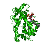
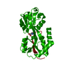
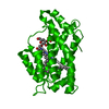
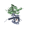
| Title | Crystal Structure of D48V mutant of Human Glycolipid Transfer Protein complexed with 3-O-sulfo galactosylceramide containing nervonoyl acyl chain | ||||||
 Components Components | Glycolipid transfer protein | ||||||
 Keywords Keywords | LIPID TRANSPORT / GLTP-fold / transport | ||||||
| Function / homology |  Function and homology information Function and homology informationGlycosphingolipid biosynthesis / glycolipid transfer activity / lipid transfer activity / ceramide 1-phosphate binding / ceramide 1-phosphate transfer activity / ER to Golgi ceramide transport / glycolipid binding / glycosphingolipid metabolic process / intermembrane lipid transfer / response to immobilization stress ...Glycosphingolipid biosynthesis / glycolipid transfer activity / lipid transfer activity / ceramide 1-phosphate binding / ceramide 1-phosphate transfer activity / ER to Golgi ceramide transport / glycolipid binding / glycosphingolipid metabolic process / intermembrane lipid transfer / response to immobilization stress / lipid binding / identical protein binding / cytosolSimilarity search - Function | ||||||
| Biological species |  Homo sapiens (human) Homo sapiens (human) | ||||||
| Method | X-RAY DIFFRACTION / SYNCHROTRON / MOLECULAR REPLACEMENT / Resolution: 1.5 Å | ||||||
 Authors Authors | Samygina, V. / Popov, A.N. / Cabo-Bilbao, A. / Ochoa-Lizarralde, B. / Patel, D.J. / Brown, R.E. / Malinina, L. | ||||||
 Citation Citation | Journal: Structure / Year: 2011 Title: Enhanced selectivity for sulfatide by engineered human glycolipid transfer protein. Authors: Samygina, V.R. / Popov, A.N. / Cabo-Bilbao, A. / Ochoa-Lizarralde, B. / Goni-de-Cerio, F. / Zhai, X. / Molotkovsky, J.G. / Patel, D.J. / Brown, R.E. / Malinina, L. | ||||||
| History |
|
- Structure visualization
| Structure viewer | Molecule: MolmilJmol/JSmol |
|---|
- Downloads & links
-Download
| PDBx/mmCIF format | 3s0i.cif.gz | 108 KB | Display | PDBx/mmCIF format |
|---|---|---|---|---|
| PDB format | pdb3s0i.ent.gz | 81.7 KB | Display | PDB format |
| PDBx/mmJSON format | 3s0i.json.gz | Tree view | PDBx/mmJSON format | |
| Others |  Other downloads Other downloads |
-Validation report
| Arichive directory | https://data.pdbj.org/pub/pdb/validation_reports/s0/3s0iftp://data.pdbj.org/pub/pdb/validation_reports/s0/3s0i | HTTPS FTP |
|---|
-Related structure data
| Related structure data |  3ricC  3rwvC  3rznSC  3s0kC S: Starting model for refinement C: citing same article ( |
|---|---|
| Similar structure data |


-Links
 PDBj
PDBj- Assembly
| Deposited unit | 
| ||||||||
|---|---|---|---|---|---|---|---|---|---|
| 1 | 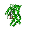
| ||||||||
| Unit cell |
|
-Components
| #1: Protein | / GLTP Mass: 23861.820 Da / Num. of mol.: 1 / Mutation: D48V Source method: isolated from a genetically manipulated source Source: (gene. exp.) Homo sapiens (human) / Gene: GLTP / Production host:  Escherichia coli (E. coli) / References: UniProt: Q9NZD2 Escherichia coli (E. coli) / References: UniProt: Q9NZD2 |
|---|---|
| #2: Chemical | ChemComp-CIS / (  Mass: 890.301 Da / Num. of mol.: 1 / Source method: obtained synthetically / Formula: C48H91NO11S Mass: 890.301 Da / Num. of mol.: 1 / Source method: obtained synthetically / Formula: C48H91NO11S |
| #3: Water | ChemComp-HOH / Water Mass: 18.015 Da / Num. of mol.: 168 / Source method: isolated from a natural source / Formula: H2O Mass: 18.015 Da / Num. of mol.: 168 / Source method: isolated from a natural source / Formula: H2O |
-Experimental details
-Experiment
| Experiment | Method: X-RAY DIFFRACTION / Number of used crystals: 1 |
|---|
- Sample preparation
| Crystal | Density Matthews: 2.01 Å3/Da / Density % sol: 38.68 % |
|---|---|
| Crystal grow | Temperature: 273 K / pH: 7 Details: 20-25% PEG 3350, pH 7.0, VAPOR DIFFUSION, HANGING DROP, temperature 273K |
-Data collection
| Diffraction | Mean temperature: 100 K |
|---|---|
| Diffraction source | Source: SYNCHROTRON / Site: ESRF  / Beamline: ID23-1 / Wavelength: 0.9754 / Beamline: ID23-1 / Wavelength: 0.9754 |
| Detector | Type: ADSC QUANTUM 315r / Detector: CCD / Date: Nov 12, 2009 |
| Radiation | Protocol: SINGLE WAVELENGTH / Monochromatic (M) / Laue (L): M / Scattering type: x-ray |
| Radiation wavelength | Wavelength: 0.9754 Å / Relative weight: 1 |
| Reflection | Resolution: 1.5→30 Å / Num. obs: 30330 / % possible obs: 99.9 % / Observed criterion σ(I): 2 / Redundancy: 5.1 % / Rmerge(I) obs: 0.061 / Net I/σ(I): 8.8 |
| Reflection shell | Resolution: 1.5→1.55 Å / Redundancy: 3.2 % / Rmerge(I) obs: 0.444 / Mean I/σ(I) obs: 2 / % possible all: 99.9 |
- Processing
| Software |
| ||||||||||||||||||||||||||||||||||||||||||||||||||||||||||||||||||||||||||||||||||||||||||||||||||||||||||||||||||||||||||||||||||||||||||||||||||||||||||||||||||||||||||
|---|---|---|---|---|---|---|---|---|---|---|---|---|---|---|---|---|---|---|---|---|---|---|---|---|---|---|---|---|---|---|---|---|---|---|---|---|---|---|---|---|---|---|---|---|---|---|---|---|---|---|---|---|---|---|---|---|---|---|---|---|---|---|---|---|---|---|---|---|---|---|---|---|---|---|---|---|---|---|---|---|---|---|---|---|---|---|---|---|---|---|---|---|---|---|---|---|---|---|---|---|---|---|---|---|---|---|---|---|---|---|---|---|---|---|---|---|---|---|---|---|---|---|---|---|---|---|---|---|---|---|---|---|---|---|---|---|---|---|---|---|---|---|---|---|---|---|---|---|---|---|---|---|---|---|---|---|---|---|---|---|---|---|---|---|---|---|---|---|---|---|---|
| Refinement | Method to determine structure: MOLECULAR REPLACEMENT Starting model: 3RZN Resolution: 1.5→15 Å / Cor.coef. Fo:Fc: 0.974 / Cor.coef. Fo:Fc free: 0.957 / SU B: 2.82 / SU ML: 0.048 / Cross valid method: THROUGHOUT / ESU R: 0.094 / ESU R Free: 0.08 / Stereochemistry target values: MAXIMUM LIKELIHOOD / Details: HYDROGENS HAVE BEEN USED IF PRESENT IN THE INPUT
| ||||||||||||||||||||||||||||||||||||||||||||||||||||||||||||||||||||||||||||||||||||||||||||||||||||||||||||||||||||||||||||||||||||||||||||||||||||||||||||||||||||||||||
| Solvent computation | Ion probe radii: 0.8 Å / Shrinkage radii: 0.8 Å / VDW probe radii: 1.2 Å / Solvent model: MASK | ||||||||||||||||||||||||||||||||||||||||||||||||||||||||||||||||||||||||||||||||||||||||||||||||||||||||||||||||||||||||||||||||||||||||||||||||||||||||||||||||||||||||||
| Displacement parameters | Biso mean: 29.447 Å2
| ||||||||||||||||||||||||||||||||||||||||||||||||||||||||||||||||||||||||||||||||||||||||||||||||||||||||||||||||||||||||||||||||||||||||||||||||||||||||||||||||||||||||||
| Refinement step | Cycle: LAST / Resolution: 1.5→15 Å
| ||||||||||||||||||||||||||||||||||||||||||||||||||||||||||||||||||||||||||||||||||||||||||||||||||||||||||||||||||||||||||||||||||||||||||||||||||||||||||||||||||||||||||
| Refine LS restraints |
| ||||||||||||||||||||||||||||||||||||||||||||||||||||||||||||||||||||||||||||||||||||||||||||||||||||||||||||||||||||||||||||||||||||||||||||||||||||||||||||||||||||||||||
| LS refinement shell | Resolution: 1.501→1.54 Å / Total num. of bins used: 20
|
