 Movie
Movie Controller


+ Open data

- Basic information
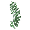
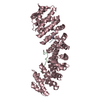
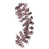
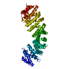
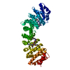
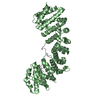
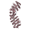
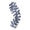
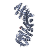
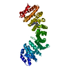
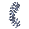
| Entry | Database: PDB / ID: 3rzx | ||||||
|---|---|---|---|---|---|---|---|
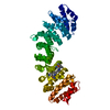
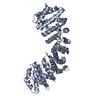
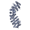
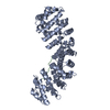
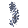
| Title | Mouse importin alpha-Ku70 NLS peptide complex | ||||||
 Components Components |
| ||||||
 Keywords Keywords |  PROTEIN TRANSPORT / Armadillo Repeats PROTEIN TRANSPORT / Armadillo Repeats | ||||||
| Function / homology |  Function and homology information Function and homology informationKu70:Ku80 complex / positive regulation of lymphocyte differentiation / DNA-dependent protein kinase complex / Sensing of DNA Double Strand Breaks / DNA-dependent protein kinase-DNA ligase 4 complex / entry of viral genome into host nucleus through nuclear pore complex via importin / cellular response to X-ray / nonhomologous end joining complex / positive regulation of viral life cycle / DNA ligation ...Ku70:Ku80 complex / positive regulation of lymphocyte differentiation / DNA-dependent protein kinase complex / Sensing of DNA Double Strand Breaks / DNA-dependent protein kinase-DNA ligase 4 complex / entry of viral genome into host nucleus through nuclear pore complex via importin / cellular response to X-ray / nonhomologous end joining complex / positive regulation of viral life cycle / DNA ligation / nuclear telomere cap complex / regulation of smooth muscle cell proliferation / Cytosolic sensors of pathogen-associated DNA / double-strand break repair via classical nonhomologous end joining / NLS-dependent protein nuclear import complex / postsynapse to nucleus signaling pathway / IRF3-mediated induction of type I IFN / host cell / recombinational repair / nuclear import signal receptor activity / nuclear localization sequence binding / cellular hyperosmotic salinity response / NLS-bearing protein import into nucleus / telomeric DNA binding / 2-LTR circle formation / Lyases; Carbon-oxygen lyases; Other carbon-oxygen lyases / 5'-deoxyribose-5-phosphate lyase activity / positive regulation of protein kinase activity / ATP-dependent activity, acting on DNA / DNA helicase activity / telomere maintenance / activation of innate immune response / cyclin binding / protein-DNA complex / Nonhomologous End-Joining (NHEJ) / Hydrolases; Acting on acid anhydrides; Acting on acid anhydrides to facilitate cellular and subcellular movement / cellular response to gamma radiation / double-strand break repair via nonhomologous end joining / cytoplasmic stress granule / protein import into nucleus / histone deacetylase binding / double-stranded DNA binding / scaffold protein binding / nuclear membrane / secretory granule lumen / DNA-binding transcription factor binding / ficolin-1-rich granule lumen / transcription regulator complex / postsynaptic density / damaged DNA binding / chromosome, telomeric region / transcription cis-regulatory region binding / innate immune response / negative regulation of DNA-templated transcription / glutamatergic synapse / Neutrophil degranulation / protein-containing complex binding / nucleolus / positive regulation of DNA-templated transcription / ATP hydrolysis activity / positive regulation of transcription by RNA polymerase II / protein-containing complex / DNA binding / RNA binding / extracellular region / nucleoplasm / ATP binding / membrane / nucleus / cytosolSimilarity search - Function | ||||||
| Biological species |  Mus musculus (house mouse) Mus musculus (house mouse) Homo sapiens (human) Homo sapiens (human) | ||||||
| Method | X-RAY DIFFRACTION / SYNCHROTRON / MOLECULAR REPLACEMENT / Resolution: 2.61 Å | ||||||
 Authors Authors | Takeda, A.A.S. / Fontes, M.R.M. | ||||||
 Citation Citation | Journal: J.Mol.Biol. / Year: 2011 Title: Structural basis of importin-alpha-mediated nuclear transport for Ku70 and Ku80. Authors: Takeda, A.A. / de Barros, A.C. / Chang, C.W. / Kobe, B. / Fontes, M.R. | ||||||
| History |
|
- Structure visualization
| Structure viewer | Molecule: MolmilJmol/JSmol |
|---|
- Downloads & links
-Download
| PDBx/mmCIF format | 3rzx.cif.gz | 99.2 KB | Display | PDBx/mmCIF format |
|---|---|---|---|---|
| PDB format | pdb3rzx.ent.gz | 73.8 KB | Display | PDB format |
| PDBx/mmJSON format | 3rzx.json.gz | Tree view | PDBx/mmJSON format | |
| Others |  Other downloads Other downloads |
-Validation report
| Arichive directory | https://data.pdbj.org/pub/pdb/validation_reports/rz/3rzxftp://data.pdbj.org/pub/pdb/validation_reports/rz/3rzx | HTTPS FTP |
|---|
-Related structure data
| Related structure data | 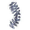 3rz9C  1pjnS S: Starting model for refinement C: citing same article ( |
|---|---|
| Similar structure data |


-Links
 PDBj
PDBj


- Assembly
| Deposited unit | 
| ||||||||
|---|---|---|---|---|---|---|---|---|---|
| 1 |
| ||||||||
| Unit cell |
|
-Components
| #1: Protein | / Importin alpha P1 / Karyopherin subunit alpha-2 / Pendulin / Pore targeting complex 58 kDa subunit ...Importin alpha P1 / Karyopherin subunit alpha-2 / Pendulin / Pore targeting complex 58 kDa subunit / PTAC58 / RAG cohort protein 1 / SRP1-alpha Mass: 55330.566 Da / Num. of mol.: 1 / Fragment: NLS binding domain Source method: isolated from a genetically manipulated source Source: (gene. exp.) Mus musculus (house mouse) / Gene: Kpna2, Rch1 / Plasmid: pET28a / Production host:  Escherichia coli (E. coli) / Strain (production host): BL21(DE3)pLysS / References: UniProt: P52293 Escherichia coli (E. coli) / Strain (production host): BL21(DE3)pLysS / References: UniProt: P52293 |
|---|---|
| #2: Protein/peptide | Mass: 2474.772 Da / Num. of mol.: 1 / Fragment: Nuclear localization signal / Source method: obtained synthetically / Source: (synth.) Homo sapiens (human) / References: UniProt: P12956 |
| #3: Water | ChemComp-HOH / Water Mass: 18.015 Da / Num. of mol.: 96 / Source method: isolated from a natural source / Formula: H2O Mass: 18.015 Da / Num. of mol.: 96 / Source method: isolated from a natural source / Formula: H2O |
-Experimental details
-Experiment
| Experiment | Method: X-RAY DIFFRACTION / Number of used crystals: 1 |
|---|
- Sample preparation
| Crystal | Density Matthews: 3.06 Å3/Da / Density % sol: 59.81 % |
|---|---|
| Crystal grow | Temperature: 293 K / Method: vapor diffusion, hanging drop / pH: 6 Details: Sodium Citrate, DTT, pH 6.0, VAPOR DIFFUSION, HANGING DROP, temperature 293K |
-Data collection
| Diffraction | Mean temperature: 100 K |
|---|---|
| Diffraction source | Source: SYNCHROTRON / Site: LNLS  / Beamline: W01B-MX2 / Wavelength: 1.45859 Å / Beamline: W01B-MX2 / Wavelength: 1.45859 Å |
| Detector | Type: MARMOSAIC 225 mm CCD / Detector: CCD / Date: Feb 27, 2008 / Details: SAGITALLY FOCUSED Si(111) |
| Radiation | Monochromator: SAGITALLY FOCUSED Si(111) / Protocol: SINGLE WAVELENGTH / Monochromatic (M) / Laue (L): M / Scattering type: x-ray |
| Radiation wavelength | Wavelength: 1.45859 Å / Relative weight: 1 |
| Reflection | Resolution: 2.6→29.07 Å / Num. all: 22152 / Num. obs: 20974 / % possible obs: 99.2 % / Observed criterion σ(F): 0 / Observed criterion σ(I): -3 |
| Reflection shell | Resolution: 2.6→2.69 Å / % possible all: 99.9 |
- Processing
| Software |
| ||||||||||||||||||||||||||||||||||||||||||||||||||||||||||||||||||||||||||||||||||||||||||||||||||||||||||||||||||||||||||||||||||||||||||||||||||||||||||||||||||||||||||
|---|---|---|---|---|---|---|---|---|---|---|---|---|---|---|---|---|---|---|---|---|---|---|---|---|---|---|---|---|---|---|---|---|---|---|---|---|---|---|---|---|---|---|---|---|---|---|---|---|---|---|---|---|---|---|---|---|---|---|---|---|---|---|---|---|---|---|---|---|---|---|---|---|---|---|---|---|---|---|---|---|---|---|---|---|---|---|---|---|---|---|---|---|---|---|---|---|---|---|---|---|---|---|---|---|---|---|---|---|---|---|---|---|---|---|---|---|---|---|---|---|---|---|---|---|---|---|---|---|---|---|---|---|---|---|---|---|---|---|---|---|---|---|---|---|---|---|---|---|---|---|---|---|---|---|---|---|---|---|---|---|---|---|---|---|---|---|---|---|---|---|---|
| Refinement | Method to determine structure: MOLECULAR REPLACEMENT Starting model: PDB ENTRY 1PJN Resolution: 2.61→29.07 Å / Cor.coef. Fo:Fc: 0.966 / Cor.coef. Fo:Fc free: 0.933 / SU B: 7.332 / SU ML: 0.157 / Cross valid method: THROUGHOUT / ESU R: 0.3 / ESU R Free: 0.238 / Stereochemistry target values: MAXIMUM LIKELIHOOD / Details: HYDROGENS HAVE BEEN ADDED IN THE RIDING POSITIONS
| ||||||||||||||||||||||||||||||||||||||||||||||||||||||||||||||||||||||||||||||||||||||||||||||||||||||||||||||||||||||||||||||||||||||||||||||||||||||||||||||||||||||||||
| Solvent computation | Ion probe radii: 0.8 Å / Shrinkage radii: 0.8 Å / VDW probe radii: 1.2 Å / Solvent model: MASK | ||||||||||||||||||||||||||||||||||||||||||||||||||||||||||||||||||||||||||||||||||||||||||||||||||||||||||||||||||||||||||||||||||||||||||||||||||||||||||||||||||||||||||
| Displacement parameters | Biso mean: 53.172 Å2
| ||||||||||||||||||||||||||||||||||||||||||||||||||||||||||||||||||||||||||||||||||||||||||||||||||||||||||||||||||||||||||||||||||||||||||||||||||||||||||||||||||||||||||
| Refinement step | Cycle: LAST / Resolution: 2.61→29.07 Å
| ||||||||||||||||||||||||||||||||||||||||||||||||||||||||||||||||||||||||||||||||||||||||||||||||||||||||||||||||||||||||||||||||||||||||||||||||||||||||||||||||||||||||||
| Refine LS restraints |
| ||||||||||||||||||||||||||||||||||||||||||||||||||||||||||||||||||||||||||||||||||||||||||||||||||||||||||||||||||||||||||||||||||||||||||||||||||||||||||||||||||||||||||
| LS refinement shell | Resolution: 2.61→2.674 Å / Total num. of bins used: 20
|
