 Movie
Movie Controller

[English] 日本語
 Yorodumi
- PDB-3qzq: Human enterovirus 71 3C protease mutant E71D in complex with rupi... -
+ Open data

- Basic information
| Entry | Database: PDB / ID: 3qzq | ||||||
|---|---|---|---|---|---|---|---|
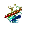
| Title | Human enterovirus 71 3C protease mutant E71D in complex with rupintrivir | ||||||
 Components Components | 3C protein | ||||||
 Keywords Keywords | HYDROLASE/HYDROLASE INHIBITOR / chymotrypsin-fold / beta-ribbon /  hydrolysis / nucleus / HYDROLASE-HYDROLASE INHIBITOR complex hydrolysis / nucleus / HYDROLASE-HYDROLASE INHIBITOR complex | ||||||
| Function / homology |  Function and homology information Function and homology informationT=pseudo3 icosahedral viral capsid / host cell cytoplasm / symbiont entry into host cell / cysteine-type endopeptidase activity / virion attachment to host cell / proteolysis / cytoplasmSimilarity search - Function | ||||||
| Biological species |   Human enterovirus 71 Human enterovirus 71 | ||||||
| Method | X-RAY DIFFRACTION / MOLECULAR REPLACEMENT / Resolution: 1.7001 Å | ||||||
 Authors Authors | Wang, J. / Fan, T. / Yao, X. / Wu, Z. / Guo, L. / Lei, X. / Wang, J. / Wang, M. / Jin, Q. / Cui, S. | ||||||
 Citation Citation | Journal: J.Virol. / Year: 2011 Title: Crystal Structures of Enterovirus 71 3C Protease Complexed with Rupintrivir Reveal the Roles of Catalytically Important Residues. Authors: Wang, J. / Fan, T. / Yao, X. / Wu, Z. / Guo, L. / Lei, X. / Wang, J. / Wang, M. / Jin, Q. / Cui, S. | ||||||
| History |
|
- Structure visualization
| Structure viewer | Molecule: MolmilJmol/JSmol |
|---|
- Downloads & links
-Download
| PDBx/mmCIF format | 3qzq.cif.gz | 447.9 KB | Display | PDBx/mmCIF format |
|---|---|---|---|---|
| PDB format | pdb3qzq.ent.gz | 377.9 KB | Display | PDB format |
| PDBx/mmJSON format | 3qzq.json.gz | Tree view | PDBx/mmJSON format | |
| Others |  Other downloads Other downloads |
-Validation report
| Arichive directory | https://data.pdbj.org/pub/pdb/validation_reports/qz/3qzqftp://data.pdbj.org/pub/pdb/validation_reports/qz/3qzq | HTTPS FTP |
|---|
-Related structure data


-Links
 PDBj
PDBj

- Assembly
| Deposited unit | 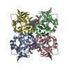
| ||||||||
|---|---|---|---|---|---|---|---|---|---|
| 1 | 
| ||||||||
| 2 | 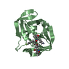
| ||||||||
| 3 | 
| ||||||||
| 4 | 
| ||||||||
| Unit cell |
| ||||||||
| Components on special symmetry positions |
|
-Components
| #1: Protein | Mass: 20588.578 Da / Num. of mol.: 4 / Mutation: E71D Source method: isolated from a genetically manipulated source Source: (gene. exp.) Human enterovirus 71 / Plasmid: pET-28a / Production host:  Escherichia coli (E. coli) / Strain (production host): Rosetta / References: UniProt: E7E815 Escherichia coli (E. coli) / Strain (production host): Rosetta / References: UniProt: E7E815#2: Chemical | ChemComp-AG7 / Rupintrivir  Mass: 600.678 Da / Num. of mol.: 4 / Source method: obtained synthetically / Formula: C31H41FN4O7 / Comment: antivirus, protease inhibitor*YM Mass: 600.678 Da / Num. of mol.: 4 / Source method: obtained synthetically / Formula: C31H41FN4O7 / Comment: antivirus, protease inhibitor*YM#3: Water | ChemComp-HOH / | Water Mass: 18.015 Da / Num. of mol.: 598 / Source method: isolated from a natural source / Formula: H2O Mass: 18.015 Da / Num. of mol.: 598 / Source method: isolated from a natural source / Formula: H2O |
|---|
-Experimental details
-Experiment
| Experiment | Method: X-RAY DIFFRACTION / Number of used crystals: 1 |
|---|
- Sample preparation
| Crystal | Density Matthews: 2.14 Å3/Da / Density % sol: 42.59 % |
|---|---|
| Crystal grow | Temperature: 295 K / Method: vapor diffusion, hanging drop / pH: 7.3 Details: Hepes 0.1M, PEG4000 10%, iso-propanol 20%, pH 7.3, VAPOR DIFFUSION, HANGING DROP, temperature 295.0K |
-Data collection
| Diffraction | Mean temperature: 100 K |
|---|---|
| Diffraction source | Source: ROTATING ANODE / Type: RIGAKU MICROMAX-007 HF / Wavelength: 1.54 Å |
| Detector | Type: RIGAKU SATURN 944+ / Detector: CCD / Date: Dec 19, 2010 |
| Radiation | Monochromator: Ge multilayers / Protocol: SINGLE WAVELENGTH / Monochromatic (M) / Laue (L): M / Scattering type: x-ray |
| Radiation wavelength | Wavelength: 1.54 Å / Relative weight: 1 |
| Reflection | Resolution: 1.7→28.49 Å / Num. obs: 73601 / % possible obs: 93.3 % / Observed criterion σ(F): 3 / Observed criterion σ(I): 3 / Redundancy: 3.18 % / Rmerge(I) obs: 0.055 / Net I/σ(I): 8.7 |
| Reflection shell | Resolution: 1.7→1.76 Å / Redundancy: 1.77 % / Rmerge(I) obs: 0.02 / Mean I/σ(I) obs: 22.7 / Num. unique all: 7772 / % possible all: 79.5 |
- Processing
| Software |
| |||||||||||||||||||||||||||||||||||||||||||||||||||||||||||||||||||||||||||||||||||||||||||||||||||||||||||||||||||||||||||||||||||||||||||||||||||||||||||||||||||||||||||||||||||||||||||||
|---|---|---|---|---|---|---|---|---|---|---|---|---|---|---|---|---|---|---|---|---|---|---|---|---|---|---|---|---|---|---|---|---|---|---|---|---|---|---|---|---|---|---|---|---|---|---|---|---|---|---|---|---|---|---|---|---|---|---|---|---|---|---|---|---|---|---|---|---|---|---|---|---|---|---|---|---|---|---|---|---|---|---|---|---|---|---|---|---|---|---|---|---|---|---|---|---|---|---|---|---|---|---|---|---|---|---|---|---|---|---|---|---|---|---|---|---|---|---|---|---|---|---|---|---|---|---|---|---|---|---|---|---|---|---|---|---|---|---|---|---|---|---|---|---|---|---|---|---|---|---|---|---|---|---|---|---|---|---|---|---|---|---|---|---|---|---|---|---|---|---|---|---|---|---|---|---|---|---|---|---|---|---|---|---|---|---|---|---|---|---|
| Refinement | Method to determine structure: MOLECULAR REPLACEMENT / Resolution: 1.7001→28.059 Å / SU ML: 0.22 / σ(F): 1.33 / Phase error: 35.39 / Stereochemistry target values: ML
| |||||||||||||||||||||||||||||||||||||||||||||||||||||||||||||||||||||||||||||||||||||||||||||||||||||||||||||||||||||||||||||||||||||||||||||||||||||||||||||||||||||||||||||||||||||||||||||
| Solvent computation | Shrinkage radii: 0.9 Å / VDW probe radii: 1.11 Å / Solvent model: FLAT BULK SOLVENT MODEL / Bsol: 40.667 Å2 / ksol: 0.404 e/Å3 | |||||||||||||||||||||||||||||||||||||||||||||||||||||||||||||||||||||||||||||||||||||||||||||||||||||||||||||||||||||||||||||||||||||||||||||||||||||||||||||||||||||||||||||||||||||||||||||
| Displacement parameters |
| |||||||||||||||||||||||||||||||||||||||||||||||||||||||||||||||||||||||||||||||||||||||||||||||||||||||||||||||||||||||||||||||||||||||||||||||||||||||||||||||||||||||||||||||||||||||||||||
| Refinement step | Cycle: LAST / Resolution: 1.7001→28.059 Å
| |||||||||||||||||||||||||||||||||||||||||||||||||||||||||||||||||||||||||||||||||||||||||||||||||||||||||||||||||||||||||||||||||||||||||||||||||||||||||||||||||||||||||||||||||||||||||||||
| Refine LS restraints |
| |||||||||||||||||||||||||||||||||||||||||||||||||||||||||||||||||||||||||||||||||||||||||||||||||||||||||||||||||||||||||||||||||||||||||||||||||||||||||||||||||||||||||||||||||||||||||||||
| LS refinement shell |
|
