 Movie
Movie Controller

[English] 日本語
 Yorodumi
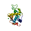
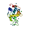
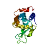
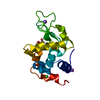
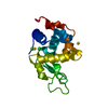
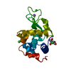
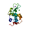
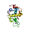
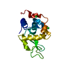
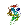
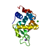
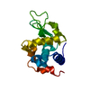
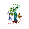
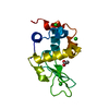
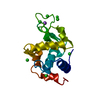
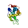
- PDB-3qe8: Crystal Structure Analysis of Lysozyme-bound fac-[Re(CO)3(H2O)(Im)]+ -
+ Open data

- Basic information
| Entry | Database: PDB / ID: 3qe8 | ||||||
|---|---|---|---|---|---|---|---|
| Title | Crystal Structure Analysis of Lysozyme-bound fac-[Re(CO)3(H2O)(Im)]+ | ||||||
 Components Components | Lysozyme C | ||||||
 Keywords Keywords |  HYDROLASE / RHENIUM COMPLEX / METALLATION HYDROLASE / RHENIUM COMPLEX / METALLATION | ||||||
| Function / homology |  Function and homology informationAntimicrobial peptides / Neutrophil degranulation / beta-N-acetylglucosaminidase activity / cell wall macromolecule catabolic process / lysozyme / lysozyme activity / killing of cells of another organism / defense response to Gram-negative bacterium / defense response to Gram-positive bacterium / defense response to bacterium ...Antimicrobial peptides / Neutrophil degranulation / beta-N-acetylglucosaminidase activity / cell wall macromolecule catabolic process / lysozyme / lysozyme activity / killing of cells of another organism / defense response to Gram-negative bacterium / defense response to Gram-positive bacterium / defense response to bacterium / endoplasmic reticulum / extracellular space / identical protein binding / cytoplasm Function and homology informationAntimicrobial peptides / Neutrophil degranulation / beta-N-acetylglucosaminidase activity / cell wall macromolecule catabolic process / lysozyme / lysozyme activity / killing of cells of another organism / defense response to Gram-negative bacterium / defense response to Gram-positive bacterium / defense response to bacterium ...Antimicrobial peptides / Neutrophil degranulation / beta-N-acetylglucosaminidase activity / cell wall macromolecule catabolic process / lysozyme / lysozyme activity / killing of cells of another organism / defense response to Gram-negative bacterium / defense response to Gram-positive bacterium / defense response to bacterium / endoplasmic reticulum / extracellular space / identical protein binding / cytoplasmSimilarity search - Function | ||||||
| Biological species |  Gallus gallus (chicken) Gallus gallus (chicken) | ||||||
| Method | X-RAY DIFFRACTION / SYNCHROTRON / MOLECULAR REPLACEMENT / Resolution: 1.49 Å | ||||||
 Authors Authors | Zobi, F. / Spingler, B. | ||||||
 Citation Citation | Journal: Inorg.Chem. / Year: 2012 Title: Post-protein-binding reactivity and modifications of the fac-[Re(CO)3]+ core Authors: Zobi, F. / Spingler, B. | ||||||
| History |
|
- Structure visualization
| Structure viewer | Molecule: MolmilJmol/JSmol |
|---|
- Downloads & links
-Download
| PDBx/mmCIF format | 3qe8.cif.gz | 68.4 KB | Display | PDBx/mmCIF format |
|---|---|---|---|---|
| PDB format | pdb3qe8.ent.gz | 49.3 KB | Display | PDB format |
| PDBx/mmJSON format | 3qe8.json.gz | Tree view | PDBx/mmJSON format | |
| Others |  Other downloads Other downloads |
-Validation report
| Arichive directory | https://data.pdbj.org/pub/pdb/validation_reports/qe/3qe8ftp://data.pdbj.org/pub/pdb/validation_reports/qe/3qe8 | HTTPS FTP |
|---|
-Related structure data
| Related structure data | 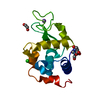 3qngC 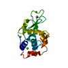 1ieeS C: citing same article ( S: Starting model for refinement |
|---|---|
| Similar structure data |


-Links
 PDBj
PDBj


- Assembly
| Deposited unit | 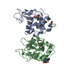
| ||||||||
|---|---|---|---|---|---|---|---|---|---|
| 1 | 
| ||||||||
| 2 | 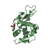
| ||||||||
| Unit cell |
|
-Components
| #1: Protein | Mass: 14331.160 Da / Num. of mol.: 2 / Source method: isolated from a natural source / Source: (natural) Gallus gallus (chicken) / References: UniProt: P00698, lysozyme#2: Chemical | 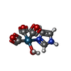  Mass: 356.330 Da / Num. of mol.: 2 / Source method: obtained synthetically / Formula: C6H6N2O4Re Mass: 356.330 Da / Num. of mol.: 2 / Source method: obtained synthetically / Formula: C6H6N2O4Re#3: Chemical | Ethylene glycol  Mass: 62.068 Da / Num. of mol.: 2 / Source method: obtained synthetically / Formula: C2H6O2 Mass: 62.068 Da / Num. of mol.: 2 / Source method: obtained synthetically / Formula: C2H6O2#4: Chemical | Chloride  Mass: 35.453 Da / Num. of mol.: 3 / Source method: obtained synthetically / Formula: Cl Mass: 35.453 Da / Num. of mol.: 3 / Source method: obtained synthetically / Formula: Cl#5: Water | ChemComp-HOH / | Water Mass: 18.015 Da / Num. of mol.: 169 / Source method: isolated from a natural source / Formula: H2O Mass: 18.015 Da / Num. of mol.: 169 / Source method: isolated from a natural source / Formula: H2O |
|---|
-Experimental details
-Experiment
| Experiment | Method: X-RAY DIFFRACTION / Number of used crystals: 1 |
|---|
- Sample preparation
| Crystal | Density Matthews: 2.01 Å3/Da / Density % sol: 38.81 % / Description: SF FILE CONTAINS FRIEDEL PAIRS |
|---|---|
| Crystal grow | Temperature: 298 K / Method: vapor diffusion, hanging drop / pH: 4.54 Details: 0.05M acetate buffer pH 4.54, 0.9M NaCl, temperature 298K, VAPOR DIFFUSION, HANGING DROP |
-Data collection
| Diffraction | Mean temperature: 100 K |
|---|---|
| Diffraction source | Source: SYNCHROTRON / Site: SLS  / Beamline: X06DA / Wavelength: 1 / Beamline: X06DA / Wavelength: 1 |
| Detector | Type: MARMOSAIC 225 mm CCD / Detector: CCD / Date: Jun 2, 2010 Details: MIRROR, BARTELS MONOCHROMATOR, DUAL CHANNEL CUT CRYSTALS, TOROIDAL MIRROR |
| Radiation | Monochromator: BARTELS MONOCHROMATOR / Protocol: SINGLE WAVELENGTH / Monochromatic (M) / Laue (L): M / Scattering type: x-ray |
| Radiation wavelength | Wavelength: 1 Å / Relative weight: 1 |
| Reflection | Resolution: 1.49→55.874 Å / Num. obs: 57107 / % possible obs: 93.8 % / Observed criterion σ(I): 2 / Redundancy: 4.06 % / Rmerge(I) obs: 0.0055 / Rsym value: 0.0361 / Net I/σ(I): 16.76 |
| Reflection shell | Resolution: 1.49→1.58 Å / Redundancy: 3.84 % / Rmerge(I) obs: 0.443 / Mean I/σ(I) obs: 3.28 / % possible all: 77.2 |
- Processing
| Software |
| |||||||||||||||||||||||||||||||||
|---|---|---|---|---|---|---|---|---|---|---|---|---|---|---|---|---|---|---|---|---|---|---|---|---|---|---|---|---|---|---|---|---|---|---|
| Refinement | Method to determine structure: MOLECULAR REPLACEMENT Starting model: PDB ENTRY 1IEE Resolution: 1.49→55.87 Å / Num. parameters: 8857 / Num. restraintsaints: 8383 / Cross valid method: FREE R / σ(F): 0 / Stereochemistry target values: Engh & Huber / Details: THE SF FILE CONTAINS FRIEDEL PAIRS.
| |||||||||||||||||||||||||||||||||
| Displacement parameters | Biso mean: 21.9583 Å2 | |||||||||||||||||||||||||||||||||
| Refine analyze | Num. disordered residues: 1 / Occupancy sum hydrogen: 0 / Occupancy sum non hydrogen: 2199.8 | |||||||||||||||||||||||||||||||||
| Refinement step | Cycle: LAST / Resolution: 1.49→55.87 Å
| |||||||||||||||||||||||||||||||||
| Refine LS restraints |
|
