 Movie
Movie Controller


+ Open data

- Basic information
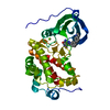
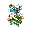
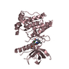
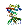
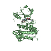
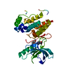
| Entry | Database: PDB / ID: 3qd2 | ||||||
|---|---|---|---|---|---|---|---|
| Title | Crystal structure of mouse PERK kinase domain | ||||||
 Components Components | Eukaryotic translation initiation factor 2-alpha kinase 3 | ||||||
 Keywords Keywords |  GENE REGULATION / eIF2a Kinase / Phosphoryalation GENE REGULATION / eIF2a Kinase / Phosphoryalation | ||||||
| Function / homology |  Function and homology information Function and homology informationPERK regulates gene expression / negative regulation of translation in response to endoplasmic reticulum stress / regulation of endoplasmic reticulum stress-induced eIF2 alpha phosphorylation / positive regulation of glutathione biosynthetic process / negative regulation of translation in response to stress / regulation of endoplasmic reticulum stress-induced intrinsic apoptotic signaling pathway / regulation of translational initiation by eIF2 alpha phosphorylation / eiF2alpha phosphorylation in response to endoplasmic reticulum stress / SREBP signaling pathway / eukaryotic initiation factor eIF2 binding ...PERK regulates gene expression / negative regulation of translation in response to endoplasmic reticulum stress / regulation of endoplasmic reticulum stress-induced eIF2 alpha phosphorylation / positive regulation of glutathione biosynthetic process / negative regulation of translation in response to stress / regulation of endoplasmic reticulum stress-induced intrinsic apoptotic signaling pathway / regulation of translational initiation by eIF2 alpha phosphorylation / eiF2alpha phosphorylation in response to endoplasmic reticulum stress / SREBP signaling pathway / eukaryotic initiation factor eIF2 binding / chondrocyte development / eukaryotic translation initiation factor 2alpha kinase activity / positive regulation of signal transduction / positive regulation of ERAD pathway / endoplasmic reticulum quality control compartment / PERK-mediated unfolded protein response / negative regulation of myelination / positive regulation of endoplasmic reticulum stress-induced intrinsic apoptotic signaling pathway / regulation of fatty acid metabolic process / endocrine pancreas development / endoplasmic reticulum organization / pancreas development / : / cellular response to cold / positive regulation of transcription by RNA polymerase I / ER overload response / fat cell differentiation / bone mineralization / intrinsic apoptotic signaling pathway in response to endoplasmic reticulum stress / positive regulation of vascular endothelial growth factor production / cellular response to glucose starvation / : / endoplasmic reticulum unfolded protein response / cellular response to amino acid starvation / lactation / response to endoplasmic reticulum stress / ossification / insulin-like growth factor receptor signaling pathway / skeletal system development / calcium-mediated signaling / Hsp90 protein binding / positive regulation of protein localization to nucleus / protein phosphatase binding / peptidyl-serine phosphorylation / angiogenesis / negative regulation of translation / protein autophosphorylation / non-specific serine/threonine protein kinase / protein kinase activity / translation / negative regulation of gene expression / protein phosphorylation / protein serine kinase activity / protein serine/threonine kinase activity / endoplasmic reticulum membrane / positive regulation of gene expression / negative regulation of apoptotic process / perinuclear region of cytoplasm / enzyme binding / endoplasmic reticulum / ATP binding / identical protein binding / nucleus / cytoplasmSimilarity search - Function | ||||||
| Biological species |  Mus musculus (house mouse) Mus musculus (house mouse) | ||||||
| Method | X-RAY DIFFRACTION / SYNCHROTRON / MOLECULAR REPLACEMENT / Resolution: 2.81 Å | ||||||
 Authors Authors | Wenjun, C. / Jingzhi, L. / David, R. / Bingdong, S. | ||||||
 Citation Citation | Journal: Acta Crystallogr.,Sect.D / Year: 2011 Title: The structure of the PERK kinase domain suggests the mechanism for its activation. Authors: Cui, W. / Li, J. / Ron, D. / Sha, B. | ||||||
| History |
|
- Structure visualization
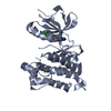
| Structure viewer | Molecule: MolmilJmol/JSmol |
|---|
- Downloads & links
-Download
| PDBx/mmCIF format | 3qd2.cif.gz | 114.4 KB | Display | PDBx/mmCIF format |
|---|---|---|---|---|
| PDB format | pdb3qd2.ent.gz | 94.2 KB | Display | PDB format |
| PDBx/mmJSON format | 3qd2.json.gz | Tree view | PDBx/mmJSON format | |
| Others |  Other downloads Other downloads |
-Validation report
| Arichive directory | https://data.pdbj.org/pub/pdb/validation_reports/qd/3qd2ftp://data.pdbj.org/pub/pdb/validation_reports/qd/3qd2 | HTTPS FTP |
|---|
-Related structure data
| Similar structure data |
|---|


-Links
 PDBj
PDBj- Assembly
| Deposited unit | 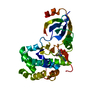
| ||||||||
|---|---|---|---|---|---|---|---|---|---|
| 1 |
| ||||||||
| Unit cell |
|
-Components
| #1: Protein | Mass: 38670.223 Da / Num. of mol.: 1 / Fragment: UNP Q9Z2B5 residues 582-701 and 867-1078 Source method: isolated from a genetically manipulated source Source: (gene. exp.) Mus musculus (house mouse) / Gene: Eif2ak3 / Production host:  Escherichia coli (E. coli) / Strain (production host): BL21 Codon Plus Escherichia coli (E. coli) / Strain (production host): BL21 Codon PlusReferences: UniProt: Q9Z2B5, non-specific serine/threonine protein kinase |
|---|---|
| #2: Water | ChemComp-HOH / Water Mass: 18.015 Da / Num. of mol.: 21 / Source method: isolated from a natural source / Formula: H2O Mass: 18.015 Da / Num. of mol.: 21 / Source method: isolated from a natural source / Formula: H2O |
| Sequence details | RESIDUES 702-866 WERE DELETED FROM UNP Q9Z2B5 |
-Experimental details
-Experiment
| Experiment | Method: X-RAY DIFFRACTION / Number of used crystals: 1 |
|---|
- Sample preparation
| Crystal | Density Matthews: 3.62 Å3/Da / Density % sol: 66.04 % |
|---|---|
| Crystal grow | Temperature: 293 K / Method: vapor diffusion, hanging drop / pH: 7.5 Details: 1.1M sodium citrate, pH 7.5, VAPOR DIFFUSION, HANGING DROP, temperature 293K |
-Data collection
| Diffraction | Mean temperature: 100 K |
|---|---|
| Diffraction source | Source: SYNCHROTRON / Site: APS  / Beamline: 22-ID / Wavelength: 0.998 Å / Beamline: 22-ID / Wavelength: 0.998 Å |
| Detector | Type: MAR CCD 165 mm / Detector: CCD / Date: Nov 3, 2009 |
| Radiation | Monochromator: insertion device / Protocol: SINGLE WAVELENGTH / Monochromatic (M) / Laue (L): M / Scattering type: x-ray |
| Radiation wavelength | Wavelength: 0.998 Å / Relative weight: 1 |
| Reflection | Resolution: 2.8→44.63 Å / Num. all: 13680 / Num. obs: 13283 / % possible obs: 97.1 % / Observed criterion σ(F): 2 / Observed criterion σ(I): 2 |
- Processing
| Software |
| ||||||||||||||||||||||||||||||||||||||||||||||||||||||||||||||||||||||||||||||||||||||||||
|---|---|---|---|---|---|---|---|---|---|---|---|---|---|---|---|---|---|---|---|---|---|---|---|---|---|---|---|---|---|---|---|---|---|---|---|---|---|---|---|---|---|---|---|---|---|---|---|---|---|---|---|---|---|---|---|---|---|---|---|---|---|---|---|---|---|---|---|---|---|---|---|---|---|---|---|---|---|---|---|---|---|---|---|---|---|---|---|---|---|---|---|
| Refinement | Method to determine structure: MOLECULAR REPLACEMENT / Resolution: 2.81→44.63 Å / Cor.coef. Fo:Fc: 0.926 / Cor.coef. Fo:Fc free: 0.896 / Occupancy max: 1 / Occupancy min: 1 / SU B: 41.669 / SU ML: 0.356 / Cross valid method: THROUGHOUT / σ(F): 0 / ESU R Free: 0.396 / Stereochemistry target values: MAXIMUM LIKELIHOOD / Details: HYDROGENS HAVE BEEN ADDED IN THE RIDING POSITIONS
| ||||||||||||||||||||||||||||||||||||||||||||||||||||||||||||||||||||||||||||||||||||||||||
| Solvent computation | Ion probe radii: 0.8 Å / Shrinkage radii: 0.8 Å / VDW probe radii: 1.2 Å / Solvent model: MASK | ||||||||||||||||||||||||||||||||||||||||||||||||||||||||||||||||||||||||||||||||||||||||||
| Displacement parameters | Biso max: 171.11 Å2 / Biso mean: 93.8811 Å2 / Biso min: 59.74 Å2
| ||||||||||||||||||||||||||||||||||||||||||||||||||||||||||||||||||||||||||||||||||||||||||
| Refinement step | Cycle: LAST / Resolution: 2.81→44.63 Å
| ||||||||||||||||||||||||||||||||||||||||||||||||||||||||||||||||||||||||||||||||||||||||||
| Refine LS restraints |
| ||||||||||||||||||||||||||||||||||||||||||||||||||||||||||||||||||||||||||||||||||||||||||
| LS refinement shell | Resolution: 2.808→2.881 Å / Total num. of bins used: 20
|
