 Movie
Movie Controller


+ Open data

- Basic information
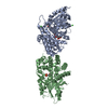
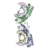
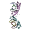
| Entry | Database: PDB / ID: 3puf | ||||||
|---|---|---|---|---|---|---|---|
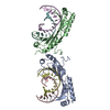
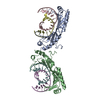
| Title | Crystal structure of human RNase H2 complex | ||||||
 Components Components |
| ||||||
 Keywords Keywords |  HYDROLASE / RNase H fold / triple barrel fold / RNase H HYDROLASE / RNase H fold / triple barrel fold / RNase H | ||||||
| Function / homology |  Function and homology information Function and homology informationribonucleotide metabolic process / ribonuclease H2 complex / DNA replication, removal of RNA primer / regulation of DNA damage checkpoint / RNA catabolic process / ribonuclease H / mismatch repair / RNA nuclease activity / regulation of G2/M transition of mitotic cell cycle / positive regulation of fibroblast proliferation ...ribonucleotide metabolic process / ribonuclease H2 complex / DNA replication, removal of RNA primer / regulation of DNA damage checkpoint / RNA catabolic process / ribonuclease H / mismatch repair / RNA nuclease activity / regulation of G2/M transition of mitotic cell cycle / positive regulation of fibroblast proliferation / RNA-DNA hybrid ribonuclease activity / gene expression / fibroblast proliferation / in utero embryonic development / DNA replication / negative regulation of gene expression / RNA binding / nucleoplasm / metal ion binding / nucleus / cytosolSimilarity search - Function | ||||||
| Biological species |  Homo sapiens (human) Homo sapiens (human) | ||||||
| Method | X-RAY DIFFRACTION / SYNCHROTRON / MOLECULAR REPLACEMENT / Resolution: 3.1 Å | ||||||
 Authors Authors | Figiel, M. / Chon, H. / Cerritelli, S.M. / Cybulska, M. / Crouch, R.J. / Nowotny, M. | ||||||
 Citation Citation | Journal: J.Biol.Chem. / Year: 2011 Title: The structural and biochemical characterization of human RNase H2 complex reveals the molecular basis for substrate recognition and Aicardi-Goutieres syndrome defects. Authors: Figiel, M. / Chon, H. / Cerritelli, S.M. / Cybulska, M. / Crouch, R.J. / Nowotny, M. | ||||||
| History |
|
- Structure visualization
| Structure viewer | Molecule: MolmilJmol/JSmol |
|---|
- Downloads & links
-Download
| PDBx/mmCIF format | 3puf.cif.gz | 650.9 KB | Display | PDBx/mmCIF format |
|---|---|---|---|---|
| PDB format | pdb3puf.ent.gz | 525.1 KB | Display | PDB format |
| PDBx/mmJSON format | 3puf.json.gz | Tree view | PDBx/mmJSON format | |
| Others |  Other downloads Other downloads |
-Validation report
| Arichive directory | https://data.pdbj.org/pub/pdb/validation_reports/pu/3pufftp://data.pdbj.org/pub/pdb/validation_reports/pu/3puf | HTTPS FTP |
|---|
-Related structure data
| Related structure data | 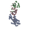 3kioS S: Starting model for refinement |
|---|---|
| Similar structure data |


-Links
 PDBj
PDBj
- Assembly
| Deposited unit | 
| ||||||||
|---|---|---|---|---|---|---|---|---|---|
| 1 | 
| ||||||||
| 2 | 
| ||||||||
| 3 | 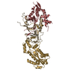
| ||||||||
| 4 | 
| ||||||||
| 5 | 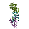
| ||||||||
| 6 | 
| ||||||||
| Unit cell |
|
-Components
| #1: Protein | Mass: 33724.156 Da / Num. of mol.: 6 Source method: isolated from a genetically manipulated source Source: (gene. exp.) Homo sapiens (human) / Gene: RNASEH2A, RNASEHI, RNHIA / Production host:  Escherichia coli (E. coli) / References: UniProt: O75792, ribonuclease H Escherichia coli (E. coli) / References: UniProt: O75792, ribonuclease H#2: Protein | Mass: 25780.783 Da / Num. of mol.: 6 Source method: isolated from a genetically manipulated source Source: (gene. exp.) Homo sapiens (human) / Gene: RNASEH2B, DLEU8 / Production host: Escherichia coli (E. coli) / References: UniProt: Q5TBB1, ribonuclease H#3: Protein | Mass: 18144.414 Da / Num. of mol.: 6 Source method: isolated from a genetically manipulated source Source: (gene. exp.) Homo sapiens (human) / Gene: RNASEH2C, AYP1 / Production host: Escherichia coli (E. coli) / References: UniProt: Q8TDP1, ribonuclease H#4: Water | ChemComp-HOH / | Water Mass: 18.015 Da / Num. of mol.: 91 / Source method: isolated from a natural source / Formula: H2O Mass: 18.015 Da / Num. of mol.: 91 / Source method: isolated from a natural source / Formula: H2O |
|---|
-Experimental details
-Experiment
| Experiment | Method: X-RAY DIFFRACTION / Number of used crystals: 1 |
|---|
- Sample preparation
| Crystal | Density Matthews: 1.99 Å3/Da / Density % sol: 38.05 % |
|---|---|
| Crystal grow | Temperature: 291 K / Method: vapor diffusion, sitting drop / pH: 5.5 Details: 0.1M MgCl2, 15% PEG 3350, 0.1M Bis-Tris (pH 5.5), and 2mM reduced glutathione, VAPOR DIFFUSION, SITTING DROP, temperature 291K |
-Data collection
| Diffraction | Mean temperature: 100 K |
|---|---|
| Diffraction source | Source: SYNCHROTRON / Site: ESRF  / Beamline: ID23-2 / Wavelength: 0.8726 Å / Beamline: ID23-2 / Wavelength: 0.8726 Å |
| Detector | Type: MARMOSAIC 225 mm CCD / Detector: CCD / Date: Sep 24, 2009 |
| Radiation | Protocol: SINGLE WAVELENGTH / Monochromatic (M) / Laue (L): M / Scattering type: x-ray |
| Radiation wavelength | Wavelength: 0.8726 Å / Relative weight: 1 |
| Reflection | Resolution: 3.1→50 Å / Num. obs: 63130 / % possible obs: 97.1 % / Observed criterion σ(F): 2 / Observed criterion σ(I): 2 / Redundancy: 2.8 % / Biso Wilson estimate: 69.09 Å2 / Rmerge(I) obs: 0.107 |
| Reflection shell | Resolution: 3.1→3.15 Å / Redundancy: 2.3 % / Rmerge(I) obs: 0.411 / Mean I/σ(I) obs: 2.2 / % possible all: 87.6 |
- Processing
| Software |
| |||||||||||||||||||||||||||||||||||||||||||||||||||||||||||||||||||||||||||||||||||||||||||||||
|---|---|---|---|---|---|---|---|---|---|---|---|---|---|---|---|---|---|---|---|---|---|---|---|---|---|---|---|---|---|---|---|---|---|---|---|---|---|---|---|---|---|---|---|---|---|---|---|---|---|---|---|---|---|---|---|---|---|---|---|---|---|---|---|---|---|---|---|---|---|---|---|---|---|---|---|---|---|---|---|---|---|---|---|---|---|---|---|---|---|---|---|---|---|---|---|---|
| Refinement | Method to determine structure: MOLECULAR REPLACEMENT Starting model: 3KIO Resolution: 3.1→44.1 Å / Cor.coef. Fo:Fc: 0.9039 / Cor.coef. Fo:Fc free: 0.8611 / Cross valid method: THROUGHOUT / σ(F): 0
| |||||||||||||||||||||||||||||||||||||||||||||||||||||||||||||||||||||||||||||||||||||||||||||||
| Displacement parameters | Biso mean: 64.38 Å2
| |||||||||||||||||||||||||||||||||||||||||||||||||||||||||||||||||||||||||||||||||||||||||||||||
| Refine analyze | Luzzati coordinate error obs: 0.524 Å | |||||||||||||||||||||||||||||||||||||||||||||||||||||||||||||||||||||||||||||||||||||||||||||||
| Refinement step | Cycle: LAST / Resolution: 3.1→44.1 Å
| |||||||||||||||||||||||||||||||||||||||||||||||||||||||||||||||||||||||||||||||||||||||||||||||
| Refine LS restraints |
| |||||||||||||||||||||||||||||||||||||||||||||||||||||||||||||||||||||||||||||||||||||||||||||||
| LS refinement shell | Resolution: 3.1→3.18 Å / Total num. of bins used: 20
|
