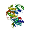
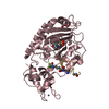
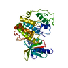
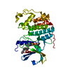
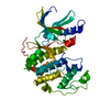
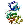
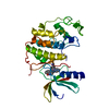
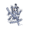
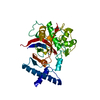
Entry Database : PDB / ID : 3prgTitle LIGAND BINDING DOMAIN OF HUMAN PEROXISOME PROLIFERATOR ACTIVATED RECEPTOR PEROXISOME PROLIFERATOR ACTIVATED RECEPTOR GAMMA Keywords / / / Function / homology Function Domain/homology Component
/ / / / / / / / / / / / / / / / / / / / / / / / / / / / / / / / / / / / / / / / / / / / / / / / / / / / / / / / / / / / / / / / / / / / / / / / / / / / / / / / / / / / / / / / / / / / / / / / / / / / / / / / / / / / / / / / / / / / / / / / / / / / / / / / / / / / / / / / Biological species Homo sapiens (human)Method / / Resolution : 2.9 Å Authors Uppenberg, J. / Svensson, C. / Jaki, M. / Bertilsson, G. / Jendeberg, L. / Berkenstam, A. Journal : J.Biol.Chem. / Year : 1998Title : Crystal structure of the ligand binding domain of the human nuclear receptor PPARgamma.Authors : Uppenberg, J. / Svensson, C. / Jaki, M. / Bertilsson, G. / Jendeberg, L. / Berkenstam, A. History Deposition Aug 24, 1998 Processing site Revision 1.0 Aug 30, 1999 Provider / Type Revision 1.1 Mar 25, 2008 Group Revision 1.2 Jul 13, 2011 Group Revision 1.3 Feb 21, 2024 Group / Database references / Category / chem_comp_bond / database_2Item / _database_2.pdbx_database_accession
Show all Show less
 Movie
Movie Controller

 Yorodumi
 Open data
 Basic information
 Components
 Keywords
 NUCLEAR RECEPTOR /
  Function and homology information
 Authors
 Citation
 Structure visualization
 Downloads & links
 Other downloads
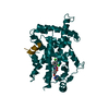
 PDBj
PDBj Assembly
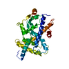

 Sample preparation
 Processing