 Movie
Movie Controller


+ Open data

- Basic information
| Entry | Database: PDB / ID: 3owt | ||||||
|---|---|---|---|---|---|---|---|
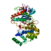
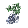
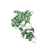
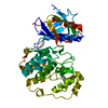
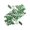
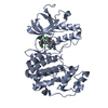
| Title | Crystal structure of S. cerevisiae RAP1-Sir3 complex | ||||||
 Components Components |
| ||||||
 Keywords Keywords |  PROTEIN BINDING / RCT domain PROTEIN BINDING / RCT domain | ||||||
| Function / homology |  Function and homology information Function and homology informationpositive regulation of ribosomal protein gene transcription by RNA polymerase II / G-quadruplex DNA formation / protection from non-homologous end joining at telomere / establishment of protein-containing complex localization to telomere / establishment of protein localization to chromatin / nuclear-transcribed mRNA catabolic process, non-stop decay / establishment of protein localization to telomere / telomere tethering at nuclear periphery / telomere maintenance via telomere lengthening / shelterin complex ...positive regulation of ribosomal protein gene transcription by RNA polymerase II / G-quadruplex DNA formation / protection from non-homologous end joining at telomere / establishment of protein-containing complex localization to telomere / establishment of protein localization to chromatin / nuclear-transcribed mRNA catabolic process, non-stop decay / establishment of protein localization to telomere / telomere tethering at nuclear periphery / telomere maintenance via telomere lengthening / shelterin complex / G-quadruplex DNA binding / double-stranded telomeric DNA binding / chromatin silencing complex / silent mating-type cassette heterochromatin formation / nucleosomal DNA binding / DNA binding, bending / regulation of glycolytic process / nuclear chromosome / telomeric DNA binding / subtelomeric heterochromatin formation / TFIID-class transcription factor complex binding / nucleosome binding / heterochromatin / cis-regulatory region sequence-specific DNA binding / heterochromatin formation / TBP-class protein binding / telomere maintenance / protein-DNA complex / double-strand break repair via nonhomologous end joining / single-stranded DNA binding / histone binding / double-stranded DNA binding / transcription regulator complex / RNA polymerase II-specific DNA-binding transcription factor binding / sequence-specific DNA binding / chromosome, telomeric region / nucleic acid binding / DNA-binding transcription factor activity, RNA polymerase II-specific / DNA-binding transcription factor activity / chromatin binding / nucleolus / regulation of DNA-templated transcription / negative regulation of transcription by RNA polymerase II / positive regulation of transcription by RNA polymerase II / mitochondrion / identical protein binding / nucleus / cytosolSimilarity search - Function | ||||||
| Biological species |  Saccharomyces cerevisiae (brewer's yeast) Saccharomyces cerevisiae (brewer's yeast) | ||||||
| Method | X-RAY DIFFRACTION / SYNCHROTRON / MOLECULAR REPLACEMENT / Resolution: 2 Å | ||||||
 Authors Authors | Chen, Y. / Yang, Y. / Lei, M. | ||||||
 Citation Citation | Journal: Nat.Struct.Mol.Biol. / Year: 2011 Title: A conserved motif within RAP1 has diversified roles in telomere protection and regulation in different organisms. Authors: Chen, Y. / Rai, R. / Zhou, Z.R. / Kanoh, J. / Ribeyre, C. / Yang, Y. / Zheng, H. / Damay, P. / Wang, F. / Tsujii, H. / Hiraoka, Y. / Shore, D. / Hu, H.Y. / Chang, S. / Lei, M. | ||||||
| History |
|
- Structure visualization
| Structure viewer | Molecule: MolmilJmol/JSmol |
|---|
- Downloads & links
-Download
| PDBx/mmCIF format | 3owt.cif.gz | 77.8 KB | Display | PDBx/mmCIF format |
|---|---|---|---|---|
| PDB format | pdb3owt.ent.gz | 58.7 KB | Display | PDB format |
| PDBx/mmJSON format | 3owt.json.gz | Tree view | PDBx/mmJSON format | |
| Others |  Other downloads Other downloads |
-Validation report
| Arichive directory | https://data.pdbj.org/pub/pdb/validation_reports/ow/3owtftp://data.pdbj.org/pub/pdb/validation_reports/ow/3owt | HTTPS FTP |
|---|
-Related structure data
| Related structure data |  2l3nC  3k6gC  3cz6S S: Starting model for refinement C: citing same article ( |
|---|---|
| Similar structure data |


-Links
 PDBj
PDBj


- Assembly
| Deposited unit | 
| ||||||||
|---|---|---|---|---|---|---|---|---|---|
| 1 |
| ||||||||
| Unit cell |
|
-Components
| #1: Protein | Mass: 18221.559 Da / Num. of mol.: 2 / Fragment: C-terminal domain (UNP residues 672 to 827) Source method: isolated from a genetically manipulated source Details: Rap1 C-terminal domain (672-827) was cloned in a PET28b-based vector with an N-terminal Sumo tag. Source: (gene. exp.) Saccharomyces cerevisiae (brewer's yeast)Gene: GRF1, N1310, RAP1, TUF1, YNL216W / Plasmid: PET28b-sumo-Rap1 / Production host:  Escherichia coli (E. coli) / Strain (production host): BL21(DE3) / References: UniProt: P11938 Escherichia coli (E. coli) / Strain (production host): BL21(DE3) / References: UniProt: P11938#2: Protein/peptide | | Mass: 3146.766 Da / Num. of mol.: 1 / Fragment: Rap1-interaction motif (UNP residues 456 to 481) Source method: isolated from a genetically manipulated source Details: Sir3 fragment (456-481) was cloned in a PET28b-based vector with an N-terminal Sumo tag. Source: (gene. exp.) Saccharomyces cerevisiae (brewer's yeast)Gene: CMT1, L9753.10, MAR2, SIR3, STE8, YLR442C / Plasmid: PET28b-sumo-Sir3 / Production host: Escherichia coli (E. coli) / Strain (production host): BL21(DE3) / References: UniProt: P06701#3: Water | ChemComp-HOH / | Water Mass: 18.015 Da / Num. of mol.: 100 / Source method: isolated from a natural source / Formula: H2O Mass: 18.015 Da / Num. of mol.: 100 / Source method: isolated from a natural source / Formula: H2O |
|---|
-Experimental details
-Experiment
| Experiment | Method: X-RAY DIFFRACTION / Number of used crystals: 1 |
|---|
- Sample preparation
| Crystal | Density Matthews: 2.08 Å3/Da / Density % sol: 40.78 % |
|---|---|
| Crystal grow | Temperature: 277 K / Method: vapor diffusion, hanging drop / pH: 4.8 Details: 100 mM sodium citrate pH 4.8, 30% PEG4K, 200 mM ammonium acetate, and 10 mM DTT , VAPOR DIFFUSION, HANGING DROP, temperature 277K |
-Data collection
| Diffraction | Mean temperature: 100 K | |||||||||||||||||||||||||||||||||
|---|---|---|---|---|---|---|---|---|---|---|---|---|---|---|---|---|---|---|---|---|---|---|---|---|---|---|---|---|---|---|---|---|---|---|
| Diffraction source | Source: SYNCHROTRON / Site: APS  / Beamline: 21-ID-D / Wavelength: 1 Å / Beamline: 21-ID-D / Wavelength: 1 Å | |||||||||||||||||||||||||||||||||
| Detector | Type: MARMOSAIC 300 mm CCD / Detector: CCD / Date: Jun 19, 2008 | |||||||||||||||||||||||||||||||||
| Radiation | Monochromator: Kohzu / Protocol: SINGLE WAVELENGTH / Monochromatic (M) / Laue (L): M / Scattering type: x-ray | |||||||||||||||||||||||||||||||||
| Radiation wavelength | Wavelength: 1 Å / Relative weight: 1 | |||||||||||||||||||||||||||||||||
| Reflection | Resolution: 1.78→50 Å / Num. obs: 27940 / % possible obs: 89.3 % / Observed criterion σ(F): 0 / Observed criterion σ(I): 0 / Redundancy: 9 % / Rmerge(I) obs: 0.064 | |||||||||||||||||||||||||||||||||
| Reflection shell |
|
- Processing
| Software |
| |||||||||||||||||||||||||||||||||||||||||||||||||||||||||||||||||||||||||||||
|---|---|---|---|---|---|---|---|---|---|---|---|---|---|---|---|---|---|---|---|---|---|---|---|---|---|---|---|---|---|---|---|---|---|---|---|---|---|---|---|---|---|---|---|---|---|---|---|---|---|---|---|---|---|---|---|---|---|---|---|---|---|---|---|---|---|---|---|---|---|---|---|---|---|---|---|---|---|---|
| Refinement | Method to determine structure: MOLECULAR REPLACEMENT Starting model: PDB entry 3CZ6 Resolution: 2→38.258 Å / SU ML: 0.31 / σ(F): 0.16 / Stereochemistry target values: ML
| |||||||||||||||||||||||||||||||||||||||||||||||||||||||||||||||||||||||||||||
| Solvent computation | Shrinkage radii: 0.9 Å / VDW probe radii: 1.11 Å / Solvent model: FLAT BULK SOLVENT MODEL / Bsol: 45.86 Å2 / ksol: 0.356 e/Å3 | |||||||||||||||||||||||||||||||||||||||||||||||||||||||||||||||||||||||||||||
| Displacement parameters |
| |||||||||||||||||||||||||||||||||||||||||||||||||||||||||||||||||||||||||||||
| Refinement step | Cycle: LAST / Resolution: 2→38.258 Å
| |||||||||||||||||||||||||||||||||||||||||||||||||||||||||||||||||||||||||||||
| Refine LS restraints |
| |||||||||||||||||||||||||||||||||||||||||||||||||||||||||||||||||||||||||||||
| LS refinement shell |
|
