 Movie
Movie Controller

[English] 日本語
 Yorodumi
- PDB-3osg: The structure of protozoan parasite Trichomonas vaginalis Myb2 in... -
+ Open data

- Basic information
| Entry | Database: PDB / ID: 3osg | ||||||
|---|---|---|---|---|---|---|---|
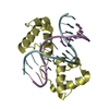
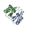
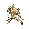
| Title | The structure of protozoan parasite Trichomonas vaginalis Myb2 in complex with MRE-1-12 DNA | ||||||
 Components Components |
| ||||||
 Keywords Keywords | transcription/DNA / transcription-DNA complex / Myb2 /  R2R3 Domain / DNA binding protein / transcription factor / Nucleus R2R3 Domain / DNA binding protein / transcription factor / Nucleus | ||||||
| Function / homology |  Function and homology information Function and homology informationDNA-binding transcription factor activity, RNA polymerase II-specific / RNA polymerase II cis-regulatory region sequence-specific DNA binding / regulation of DNA-templated transcription / nucleusSimilarity search - Function | ||||||
| Biological species |  Trichomonas vaginalis (eukaryote) Trichomonas vaginalis (eukaryote) | ||||||
| Method | X-RAY DIFFRACTION / SYNCHROTRON / MOLECULAR REPLACEMENT / Resolution: 1.997 Å | ||||||
 Authors Authors | Jiang, I. / Tsai, C.K. / Chen, S.C. / Wang, S.H. / Amiraslanov, I. / Chang, C.F. / Wu, W.J. / Tai, J.H. / Liaw, Y.C. / Huang, T.H. | ||||||
 Citation Citation | Journal: Nucleic Acids Res. / Year: 2011 Title: Molecular basis of the recognition of the ap65-1 gene transcription promoter elements by a Myb protein from the protozoan parasite Trichomonas vaginalis. Authors: Jiang, I. / Tsai, C.K. / Chen, S.C. / Wang, S.H. / Amiraslanov, I. / Chang, C.F. / Wu, W.J. / Tai, J.H. / Liaw, Y.C. / Huang, T.H. | ||||||
| History |
|
- Structure visualization
| Structure viewer | Molecule: MolmilJmol/JSmol |
|---|
- Downloads & links
-Download
| PDBx/mmCIF format | 3osg.cif.gz | 90.4 KB | Display | PDBx/mmCIF format |
|---|---|---|---|---|
| PDB format | pdb3osg.ent.gz | 65.1 KB | Display | PDB format |
| PDBx/mmJSON format | 3osg.json.gz | Tree view | PDBx/mmJSON format | |
| Others |  Other downloads Other downloads |
-Validation report
| Arichive directory | https://data.pdbj.org/pub/pdb/validation_reports/os/3osgftp://data.pdbj.org/pub/pdb/validation_reports/os/3osg | HTTPS FTP |
|---|
-Related structure data


-Links
 PDBj
PDBj


- Assembly
| Deposited unit | 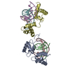
| ||||||||
|---|---|---|---|---|---|---|---|---|---|
| 1 | 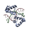
| ||||||||
| 2 | 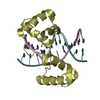
| ||||||||
| Unit cell |
|
-Components
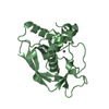
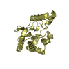
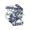
| #1: Protein | Mass: 14885.064 Da / Num. of mol.: 2 / Fragment: Myb2 R2R3 Domain Source method: isolated from a genetically manipulated source Source: (gene. exp.) Trichomonas vaginalis (eukaryote) / Gene: TVAG_211210 / Plasmid: pET-22b(+) / Production host:  Escherichia coli (E. coli) / Strain (production host): BL21(de3) / References: UniProt: Q58HP3 Escherichia coli (E. coli) / Strain (production host): BL21(de3) / References: UniProt: Q58HP3#2: DNA chain | Mass: 3660.428 Da / Num. of mol.: 2 / Source method: obtained synthetically #3: DNA chain | Mass: 3660.428 Da / Num. of mol.: 2 / Source method: obtained synthetically #4: Water | ChemComp-HOH / | Water Mass: 18.015 Da / Num. of mol.: 300 / Source method: isolated from a natural source / Formula: H2O Mass: 18.015 Da / Num. of mol.: 300 / Source method: isolated from a natural source / Formula: H2O |
|---|
-Experimental details
-Experiment
| Experiment | Method: X-RAY DIFFRACTION / Number of used crystals: 1 |
|---|
- Sample preparation
| Crystal | Density Matthews: 2.35 Å3/Da / Density % sol: 47.68 % |
|---|---|
| Crystal grow | Temperature: 298 K / Method: vapor diffusion, hanging drop / pH: 7 Details: 0.1M HEPES, 1.5-1.7M ammonium sulfate, pH 7.0, VAPOR DIFFUSION, HANGING DROP, temperature 298K |
-Data collection
| Diffraction | Mean temperature: 100 K | |||||||||||||||||||||||||||||||||
|---|---|---|---|---|---|---|---|---|---|---|---|---|---|---|---|---|---|---|---|---|---|---|---|---|---|---|---|---|---|---|---|---|---|---|
| Diffraction source | Source: SYNCHROTRON / Site: NSRRC  / Beamline: BL13B1 / Wavelength: 0.97622 Å / Beamline: BL13B1 / Wavelength: 0.97622 Å | |||||||||||||||||||||||||||||||||
| Detector | Type: ADSC QUANTUM 315 / Detector: CCD / Date: Dec 6, 2008 | |||||||||||||||||||||||||||||||||
| Radiation | Monochromator: LN2-cooled fixed-exit double crystl Si(111) monochromator Protocol: MAD / Monochromatic (M) / Laue (L): M / Scattering type: x-ray | |||||||||||||||||||||||||||||||||
| Radiation wavelength | Wavelength: 0.97622 Å / Relative weight: 1 | |||||||||||||||||||||||||||||||||
| Reflection | Resolution: 1.997→30 Å / Num. all: 27657 / Num. obs: 26911 / % possible obs: 97.3 % / Observed criterion σ(F): 1 / Observed criterion σ(I): 1 | |||||||||||||||||||||||||||||||||
| Reflection shell |
|
- Processing
| Software |
| ||||||||||||||||||||||||||||||||||||||||||||||||||||||||||||
|---|---|---|---|---|---|---|---|---|---|---|---|---|---|---|---|---|---|---|---|---|---|---|---|---|---|---|---|---|---|---|---|---|---|---|---|---|---|---|---|---|---|---|---|---|---|---|---|---|---|---|---|---|---|---|---|---|---|---|---|---|---|
| Refinement | Method to determine structure: MOLECULAR REPLACEMENT / Resolution: 1.997→24.193 Å / SU ML: 0.31 / σ(F): 0.09 / Stereochemistry target values: ML
| ||||||||||||||||||||||||||||||||||||||||||||||||||||||||||||
| Solvent computation | Shrinkage radii: 0.9 Å / VDW probe radii: 1.11 Å / Solvent model: FLAT BULK SOLVENT MODEL / Bsol: 42.221 Å2 / ksol: 0.363 e/Å3 | ||||||||||||||||||||||||||||||||||||||||||||||||||||||||||||
| Displacement parameters |
| ||||||||||||||||||||||||||||||||||||||||||||||||||||||||||||
| Refinement step | Cycle: LAST / Resolution: 1.997→24.193 Å
| ||||||||||||||||||||||||||||||||||||||||||||||||||||||||||||
| Refine LS restraints |
| ||||||||||||||||||||||||||||||||||||||||||||||||||||||||||||
| LS refinement shell | Refine-ID: X-RAY DIFFRACTION / Total num. of bins used: 9
|
