 Movie
Movie Controller

[English] 日本語
 Yorodumi
- PDB-3o2n: X-ray Crystallographic Structure Activity Relationship (SAR) of C... -
+ Open data

- Basic information
| Entry | Database: PDB / ID: 3o2n | ||||||
|---|---|---|---|---|---|---|---|
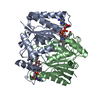
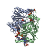
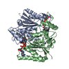
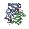
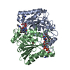
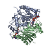
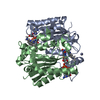
| Title | X-ray Crystallographic Structure Activity Relationship (SAR) of Casimiroin and its Analogs Bound to Human Quinone Reductase 2 | ||||||
 Components Components | Ribosyldihydronicotinamide dehydrogenase [quinone] | ||||||
 Keywords Keywords |  OXIDOREDUCTASE / protein dimer OXIDOREDUCTASE / protein dimer | ||||||
| Function / homology |  Function and homology informationribosyldihydronicotinamide dehydrogenase (quinone) / dihydronicotinamide riboside quinone reductase activity / quinone catabolic process / resveratrol binding / oxidoreductase activity, acting on other nitrogenous compounds as donors / melatonin binding / NAD(P)H dehydrogenase (quinone) activity / Phase I - Functionalization of compounds / chloride ion binding / FAD binding ...ribosyldihydronicotinamide dehydrogenase (quinone) / dihydronicotinamide riboside quinone reductase activity / quinone catabolic process / resveratrol binding / oxidoreductase activity, acting on other nitrogenous compounds as donors / melatonin binding / NAD(P)H dehydrogenase (quinone) activity / Phase I - Functionalization of compounds / chloride ion binding / FAD binding / electron transfer activity / oxidoreductase activity / protein homodimerization activity / extracellular exosome / zinc ion binding / nucleoplasm / cytosol Function and homology informationribosyldihydronicotinamide dehydrogenase (quinone) / dihydronicotinamide riboside quinone reductase activity / quinone catabolic process / resveratrol binding / oxidoreductase activity, acting on other nitrogenous compounds as donors / melatonin binding / NAD(P)H dehydrogenase (quinone) activity / Phase I - Functionalization of compounds / chloride ion binding / FAD binding ...ribosyldihydronicotinamide dehydrogenase (quinone) / dihydronicotinamide riboside quinone reductase activity / quinone catabolic process / resveratrol binding / oxidoreductase activity, acting on other nitrogenous compounds as donors / melatonin binding / NAD(P)H dehydrogenase (quinone) activity / Phase I - Functionalization of compounds / chloride ion binding / FAD binding / electron transfer activity / oxidoreductase activity / protein homodimerization activity / extracellular exosome / zinc ion binding / nucleoplasm / cytosolSimilarity search - Function | ||||||
| Biological species |  Homo sapiens (human) Homo sapiens (human) | ||||||
| Method | X-RAY DIFFRACTION / SYNCHROTRON / MOLECULAR REPLACEMENT / Resolution: 1.6 Å | ||||||
 Authors Authors | Sturdy, M. | ||||||
 Citation Citation | Journal: To be Published Title: X-ray Crystallographic Structure Activity Relationship (SAR) of Casimiroin and its Analogs Bound to Human Quinone Reductase 2 Authors: Sturdy, M. #1: Journal: J.Med.Chem. / Year: 2009Title: Synthesis of casimiroin and optimization of its quinone reductase 2 and aromatase inhibitory activities. Authors: Maiti, A. / Reddy, P.V. / Sturdy, M. / Marler, L. / Pegan, S.D. / Mesecar, A.D. / Pezzuto, J.M. / Cushman, M. | ||||||
| History |
|
- Structure visualization
| Structure viewer | Molecule: MolmilJmol/JSmol |
|---|
- Downloads & links
-Download
| PDBx/mmCIF format | 3o2n.cif.gz | 119.5 KB | Display | PDBx/mmCIF format |
|---|---|---|---|---|
| PDB format | pdb3o2n.ent.gz | 90.3 KB | Display | PDB format |
| PDBx/mmJSON format | 3o2n.json.gz | Tree view | PDBx/mmJSON format | |
| Others |  Other downloads Other downloads |
-Validation report
| Arichive directory | https://data.pdbj.org/pub/pdb/validation_reports/o2/3o2nftp://data.pdbj.org/pub/pdb/validation_reports/o2/3o2n | HTTPS FTP |
|---|
-Related structure data
| Related structure data |  3nfrC  3nhfC  3nhjC  3nhkC  3nhlC  3nhpC  3nhrC 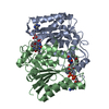 3nhsC  3nhuC  3nhwC  3nhyC  1sg0S S: Starting model for refinement C: citing same article ( |
|---|---|
| Similar structure data |


-Links
 PDBj
PDBj- Assembly
| Deposited unit | 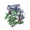
| ||||||||
|---|---|---|---|---|---|---|---|---|---|
| 1 |
| ||||||||
| Unit cell |
|
-Components
| #1: Protein | Mass: 25849.338 Da / Num. of mol.: 2 Source method: isolated from a genetically manipulated source Source: (gene. exp.) Homo sapiens (human) / Gene: NQO2, NMOR2 / Plasmid: pET-23d / Production host:  Escherichia coli (E. coli) / Strain (production host): BL21(DE3) / References: UniProt: P16083, EC: 1.10.99.2 Escherichia coli (E. coli) / Strain (production host): BL21(DE3) / References: UniProt: P16083, EC: 1.10.99.2#2: Chemical |   Mass: 65.409 Da / Num. of mol.: 2 / Source method: obtained synthetically / Formula: Zn Mass: 65.409 Da / Num. of mol.: 2 / Source method: obtained synthetically / Formula: Zn#3: Chemical | Flavin adenine dinucleotide  Mass: 785.550 Da / Num. of mol.: 2 / Source method: obtained synthetically / Formula: C27H33N9O15P2 / Comment: FAD*YM Mass: 785.550 Da / Num. of mol.: 2 / Source method: obtained synthetically / Formula: C27H33N9O15P2 / Comment: FAD*YM#4: Chemical |   Mass: 219.237 Da / Num. of mol.: 3 / Source method: obtained synthetically / Formula: C12H13NO3 Mass: 219.237 Da / Num. of mol.: 3 / Source method: obtained synthetically / Formula: C12H13NO3#5: Water | ChemComp-HOH / | Water Mass: 18.015 Da / Num. of mol.: 396 / Source method: isolated from a natural source / Formula: H2O Mass: 18.015 Da / Num. of mol.: 396 / Source method: isolated from a natural source / Formula: H2O |
|---|
-Experimental details
-Experiment
| Experiment | Method: X-RAY DIFFRACTION / Number of used crystals: 1 |
|---|
- Sample preparation
| Crystal | Density Matthews: 2.43 Å3/Da / Density % sol: 49.39 % |
|---|---|
| Crystal grow | Temperature: 298 K / Method: vapor diffusion, hanging drop / pH: 6.7 Details: 1.339 M ammonium sulfate, 0.1 M Bis-Tris, 0.1 M NaCl, 5 mM DTT, 12 uM FAD, pH 6.7, VAPOR DIFFUSION, HANGING DROP, temperature 298K |
-Data collection
| Diffraction | Mean temperature: 100 K | |||||||||||||||||||||||||||||||||||||||||||||||||||||||||||||||||||||||||||||||||||||||||||||||||||||||||||||||||||||||||||||||||||||||||||||||||||
|---|---|---|---|---|---|---|---|---|---|---|---|---|---|---|---|---|---|---|---|---|---|---|---|---|---|---|---|---|---|---|---|---|---|---|---|---|---|---|---|---|---|---|---|---|---|---|---|---|---|---|---|---|---|---|---|---|---|---|---|---|---|---|---|---|---|---|---|---|---|---|---|---|---|---|---|---|---|---|---|---|---|---|---|---|---|---|---|---|---|---|---|---|---|---|---|---|---|---|---|---|---|---|---|---|---|---|---|---|---|---|---|---|---|---|---|---|---|---|---|---|---|---|---|---|---|---|---|---|---|---|---|---|---|---|---|---|---|---|---|---|---|---|---|---|---|---|---|---|
| Diffraction source | Source: SYNCHROTRON / Site: APS  / Beamline: 21-ID-D / Wavelength: 1 Å / Beamline: 21-ID-D / Wavelength: 1 Å | |||||||||||||||||||||||||||||||||||||||||||||||||||||||||||||||||||||||||||||||||||||||||||||||||||||||||||||||||||||||||||||||||||||||||||||||||||
| Detector | Type: MARMOSAIC 300 mm CCD / Detector: CCD / Date: Jul 19, 2010 | |||||||||||||||||||||||||||||||||||||||||||||||||||||||||||||||||||||||||||||||||||||||||||||||||||||||||||||||||||||||||||||||||||||||||||||||||||
| Radiation | Monochromator: Si(111) / Protocol: SINGLE WAVELENGTH / Monochromatic (M) / Laue (L): M / Scattering type: x-ray | |||||||||||||||||||||||||||||||||||||||||||||||||||||||||||||||||||||||||||||||||||||||||||||||||||||||||||||||||||||||||||||||||||||||||||||||||||
| Radiation wavelength | Wavelength: 1 Å / Relative weight: 1 | |||||||||||||||||||||||||||||||||||||||||||||||||||||||||||||||||||||||||||||||||||||||||||||||||||||||||||||||||||||||||||||||||||||||||||||||||||
| Reflection | Resolution: 1.6→65.76 Å / Num. obs: 66255 / % possible obs: 98.7 % / Observed criterion σ(F): 0 / Observed criterion σ(I): 1 / Redundancy: 5.9 % / Rmerge(I) obs: 0.054 / Χ2: 1.238 / Net I/σ(I): 11.5 | |||||||||||||||||||||||||||||||||||||||||||||||||||||||||||||||||||||||||||||||||||||||||||||||||||||||||||||||||||||||||||||||||||||||||||||||||||
| Reflection shell |
|
- Processing
| Software |
| |||||||||||||||||||||||||||||||||||||||||||||||||||||||||||||||||
|---|---|---|---|---|---|---|---|---|---|---|---|---|---|---|---|---|---|---|---|---|---|---|---|---|---|---|---|---|---|---|---|---|---|---|---|---|---|---|---|---|---|---|---|---|---|---|---|---|---|---|---|---|---|---|---|---|---|---|---|---|---|---|---|---|---|---|
| Refinement | Method to determine structure: MOLECULAR REPLACEMENT Starting model: pdb entry 1SG0 with the ligand manually removed Resolution: 1.6→50 Å / Cor.coef. Fo:Fc: 0.966 / Cor.coef. Fo:Fc free: 0.952 / WRfactor Rfree: 0.2162 / WRfactor Rwork: 0.1771 / Occupancy max: 1 / Occupancy min: 0.5 / FOM work R set: 0.8462 / SU B: 1.832 / SU ML: 0.064 / SU R Cruickshank DPI: 0.0902 / SU Rfree: 0.0933 / Cross valid method: THROUGHOUT / σ(F): 0 / σ(I): 1 / ESU R Free: 0.093 / Stereochemistry target values: MAXIMUM LIKELIHOOD Details: HYDROGENS HAVE BEEN ADDED IN THE RIDING POSITIONS. U VALUES REFINED INDIVIDUALLY.
| |||||||||||||||||||||||||||||||||||||||||||||||||||||||||||||||||
| Solvent computation | Ion probe radii: 0.8 Å / Shrinkage radii: 0.8 Å / VDW probe radii: 1.4 Å / Solvent model: MASK | |||||||||||||||||||||||||||||||||||||||||||||||||||||||||||||||||
| Displacement parameters | Biso max: 72.92 Å2 / Biso mean: 23.3804 Å2 / Biso min: 6.49 Å2
| |||||||||||||||||||||||||||||||||||||||||||||||||||||||||||||||||
| Refinement step | Cycle: LAST / Resolution: 1.6→50 Å
| |||||||||||||||||||||||||||||||||||||||||||||||||||||||||||||||||
| Refine LS restraints |
| |||||||||||||||||||||||||||||||||||||||||||||||||||||||||||||||||
| LS refinement shell | Resolution: 1.6→1.642 Å / Total num. of bins used: 20
|
