 Movie
Movie Controller

[English] 日本語
 Yorodumi
- PDB-3nsw: Crystal Structure of Ancylostoma ceylanicum Excretory-Secretory P... -
+ Open data

- Basic information
| Entry | Database: PDB / ID: 3nsw | ||||||
|---|---|---|---|---|---|---|---|
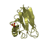
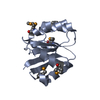
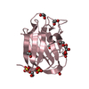
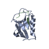
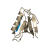
| Title | Crystal Structure of Ancylostoma ceylanicum Excretory-Secretory Protein 2 | ||||||
 Components Components | Excretory-secretory protein 2 | ||||||
 Keywords Keywords |  IMMUNE SYSTEM / Ancylostoma ceylanicum / hookworm / excretory-secretory protein / merohedral twinning / immunomodulator / netrin domain IMMUNE SYSTEM / Ancylostoma ceylanicum / hookworm / excretory-secretory protein / merohedral twinning / immunomodulator / netrin domain | ||||||
| Function / homology | OB fold (Dihydrolipoamide Acetyltransferase, E2P) - #780 / OB fold (Dihydrolipoamide Acetyltransferase, E2P) / Beta Barrel / Mainly Beta / Excretory-secretory protein 2 Function and homology information Function and homology information | ||||||
| Biological species |  Ancylostoma ceylanicum (invertebrata) Ancylostoma ceylanicum (invertebrata) | ||||||
| Method | X-RAY DIFFRACTION / SYNCHROTRON / MOLECULAR REPLACEMENT / Resolution: 1.75 Å | ||||||
 Authors Authors | Kucera, K. / Modis, Y. | ||||||
 Citation Citation | Journal: J.Mol.Biol. / Year: 2011 Title: Ancylostoma ceylanicum Excretory-Secretory Protein 2 Adopts a Netrin-Like Fold and Defines a Novel Family of Nematode Proteins. Authors: Kucera, K. / Harrison, L.M. / Cappello, M. / Modis, Y. | ||||||
| History |
|
- Structure visualization
| Structure viewer | Molecule: MolmilJmol/JSmol |
|---|
- Downloads & links
-Download
| PDBx/mmCIF format | 3nsw.cif.gz | 329 KB | Display | PDBx/mmCIF format |
|---|---|---|---|---|
| PDB format | pdb3nsw.ent.gz | 268.3 KB | Display | PDB format |
| PDBx/mmJSON format | 3nsw.json.gz | Tree view | PDBx/mmJSON format | |
| Others |  Other downloads Other downloads |
-Validation report
| Arichive directory | https://data.pdbj.org/pub/pdb/validation_reports/ns/3nswftp://data.pdbj.org/pub/pdb/validation_reports/ns/3nsw | HTTPS FTP |
|---|
-Related structure data
| Similar structure data |
|---|


-Links
 PDBj
PDBj- Assembly
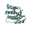
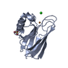
| Deposited unit | 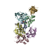
| ||||||||
|---|---|---|---|---|---|---|---|---|---|
| 1 | 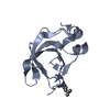
| ||||||||
| 2 | 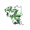
| ||||||||
| 3 | 
| ||||||||
| 4 | 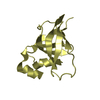
| ||||||||
| 5 | 
| ||||||||
| 6 | 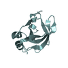
| ||||||||
| 7 | 
| ||||||||
| Unit cell |
|
-Components
| #1: Protein | Mass: 12083.514 Da / Num. of mol.: 7 Source method: isolated from a genetically manipulated source Details: Expressed with an N-terminal 6x histidine tag and thrombin cleavage sequence. Thrombin cleavage prior to crystallization left non-native Gly-Ser-His in the crystallization construct. Source: (gene. exp.) Ancylostoma ceylanicum (invertebrata) / Gene: ES-2 / Plasmid: pET28b / Production host:  Escherichia coli (E. coli) / Strain (production host): Origami2(DE3) / References: UniProt: Q6R7N7 Escherichia coli (E. coli) / Strain (production host): Origami2(DE3) / References: UniProt: Q6R7N7#2: Chemical | ChemComp-MPD / ( | 2-Methyl-2,4-pentanediol  Mass: 118.174 Da / Num. of mol.: 1 / Source method: obtained synthetically / Formula: C6H14O2 / Comment: precipitant*YM Mass: 118.174 Da / Num. of mol.: 1 / Source method: obtained synthetically / Formula: C6H14O2 / Comment: precipitant*YM#3: Chemical | ChemComp-EPE / | HEPES  Mass: 238.305 Da / Num. of mol.: 1 / Source method: obtained synthetically / Formula: C8H18N2O4S / Comment: pH buffer*YM Mass: 238.305 Da / Num. of mol.: 1 / Source method: obtained synthetically / Formula: C8H18N2O4S / Comment: pH buffer*YM#4: Water | ChemComp-HOH / | Water Mass: 18.015 Da / Num. of mol.: 928 / Source method: isolated from a natural source / Formula: H2O Mass: 18.015 Da / Num. of mol.: 928 / Source method: isolated from a natural source / Formula: H2O |
|---|
-Experimental details
-Experiment
| Experiment | Method: X-RAY DIFFRACTION / Number of used crystals: 1 |
|---|
- Sample preparation
| Crystal | Density Matthews: 2.78 Å3/Da / Density % sol: 55.79 % |
|---|---|
| Crystal grow | Temperature: 277 K / Method: vapor diffusion, hanging drop / pH: 6.8 Details: 0.1 M 2-[4-(2-hydroxyethyl)piperazin-1-yl]ethanesulfonic acid (HEPES), 60% (4S)-2-Methyl-2,4-pentanediol (MPD), pH 6.8, VAPOR DIFFUSION, HANGING DROP, temperature 277K |
-Data collection
| Diffraction | Mean temperature: 100 K | |||||||||||||||
|---|---|---|---|---|---|---|---|---|---|---|---|---|---|---|---|---|
| Diffraction source | Source: SYNCHROTRON / Site: NSLS  / Beamline: X25 / Wavelength: 1.0809 Å / Beamline: X25 / Wavelength: 1.0809 Å | |||||||||||||||
| Detector | Type: ADSC QUANTUM 315 / Detector: CCD / Date: Jul 28, 2009 Details: Meridionally-bent fused silica mirror with palladium and uncoated stripes vertically-focusing at 6.6:1 demagnification. | |||||||||||||||
| Radiation | Monochromator: Double silicon(111) crystal monochromator with cryogenically-cooled first crystal and sagittally-bent second crystal horizontally-focusing at 3.3:1 demagnification. Protocol: SINGLE WAVELENGTH / Monochromatic (M) / Laue (L): M / Scattering type: x-ray | |||||||||||||||
| Radiation wavelength | Wavelength: 1.0809 Å / Relative weight: 1 | |||||||||||||||
| Reflection twin |
| |||||||||||||||
| Reflection | Resolution: 1.75→36.91 Å / Num. obs: 79362 / % possible obs: 90.5 % / Redundancy: 4.6 % / Rmerge(I) obs: 0.074 / Net I/σ(I): 18.498 | |||||||||||||||
| Reflection shell | Resolution: 1.75→1.81 Å / Redundancy: 1.9 % / Rmerge(I) obs: 0.354 / Mean I/σ(I) obs: 2.326 / Num. unique all: 3110 / % possible all: 52.3 |
- Processing
| Software |
| ||||||||||||||||||||||||||||||||||||||||||||||||||||||||||||||||||||||||||||||||||||||||||||||||||||||||||||||||||||||||||||||||||||||||||||||||||||||||||||||||||||||||||||||||||||||||||||||||||||||||
|---|---|---|---|---|---|---|---|---|---|---|---|---|---|---|---|---|---|---|---|---|---|---|---|---|---|---|---|---|---|---|---|---|---|---|---|---|---|---|---|---|---|---|---|---|---|---|---|---|---|---|---|---|---|---|---|---|---|---|---|---|---|---|---|---|---|---|---|---|---|---|---|---|---|---|---|---|---|---|---|---|---|---|---|---|---|---|---|---|---|---|---|---|---|---|---|---|---|---|---|---|---|---|---|---|---|---|---|---|---|---|---|---|---|---|---|---|---|---|---|---|---|---|---|---|---|---|---|---|---|---|---|---|---|---|---|---|---|---|---|---|---|---|---|---|---|---|---|---|---|---|---|---|---|---|---|---|---|---|---|---|---|---|---|---|---|---|---|---|---|---|---|---|---|---|---|---|---|---|---|---|---|---|---|---|---|---|---|---|---|---|---|---|---|---|---|---|---|---|---|---|---|
| Refinement | Method to determine structure: MOLECULAR REPLACEMENT Starting model: STARTING MODEL: THE STRUCTURE OF ACEES-2 WAS SOLVED USING A LEAD (PBNO3) SOAKED CRYSTAL AND SAD METHODS AT 2.0 ANGSTROM RESOLUTION Resolution: 1.75→36.91 Å / Cor.coef. Fo:Fc: 0.969 / Cor.coef. Fo:Fc free: 0.954 / SU B: 2.981 / SU ML: 0.042 / Isotropic thermal model: Isotropic / Cross valid method: THROUGHOUT / ESU R Free: 0.02 / Stereochemistry target values: MAXIMUM LIKELIHOOD Details: Used amplitude-based twin refinement with twin operator: H, K, L fraction = 0.759 and -H, K, -L fraction = 0.241.
| ||||||||||||||||||||||||||||||||||||||||||||||||||||||||||||||||||||||||||||||||||||||||||||||||||||||||||||||||||||||||||||||||||||||||||||||||||||||||||||||||||||||||||||||||||||||||||||||||||||||||
| Solvent computation | Ion probe radii: 0.8 Å / Shrinkage radii: 0.8 Å / VDW probe radii: 1.2 Å / Solvent model: MASK | ||||||||||||||||||||||||||||||||||||||||||||||||||||||||||||||||||||||||||||||||||||||||||||||||||||||||||||||||||||||||||||||||||||||||||||||||||||||||||||||||||||||||||||||||||||||||||||||||||||||||
| Displacement parameters | Biso mean: 35.251 Å2
| ||||||||||||||||||||||||||||||||||||||||||||||||||||||||||||||||||||||||||||||||||||||||||||||||||||||||||||||||||||||||||||||||||||||||||||||||||||||||||||||||||||||||||||||||||||||||||||||||||||||||
| Refine analyze |
| ||||||||||||||||||||||||||||||||||||||||||||||||||||||||||||||||||||||||||||||||||||||||||||||||||||||||||||||||||||||||||||||||||||||||||||||||||||||||||||||||||||||||||||||||||||||||||||||||||||||||
| Refinement step | Cycle: LAST / Resolution: 1.75→36.91 Å
| ||||||||||||||||||||||||||||||||||||||||||||||||||||||||||||||||||||||||||||||||||||||||||||||||||||||||||||||||||||||||||||||||||||||||||||||||||||||||||||||||||||||||||||||||||||||||||||||||||||||||
| Refine LS restraints |
| ||||||||||||||||||||||||||||||||||||||||||||||||||||||||||||||||||||||||||||||||||||||||||||||||||||||||||||||||||||||||||||||||||||||||||||||||||||||||||||||||||||||||||||||||||||||||||||||||||||||||
| LS refinement shell | Resolution: 1.75→1.795 Å / Total num. of bins used: 20
| ||||||||||||||||||||||||||||||||||||||||||||||||||||||||||||||||||||||||||||||||||||||||||||||||||||||||||||||||||||||||||||||||||||||||||||||||||||||||||||||||||||||||||||||||||||||||||||||||||||||||
| Refinement TLS params. | Method: refined / Refine-ID: X-RAY DIFFRACTION
| ||||||||||||||||||||||||||||||||||||||||||||||||||||||||||||||||||||||||||||||||||||||||||||||||||||||||||||||||||||||||||||||||||||||||||||||||||||||||||||||||||||||||||||||||||||||||||||||||||||||||
| Refinement TLS group |
|
