- PDB-3n3x: Crystal Structure of the complex formed between type I ribosome i... -
+
Open data
ID or keywords:
Loading...
-
Basic information
Entry
Database: PDB / ID: 3n3x
Title
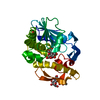
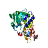
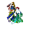
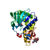
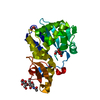
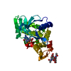
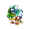
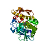
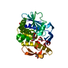
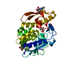
Crystal Structure of the complex formed between type I ribosome inactivating protein and hexapeptide Ser-Asp-Asp-Asp-Met-Gly at 1.7 A resolution
Components
Ribosome inactivating proteinRibosome-inactivating protein
SDDDMG peptide
Keywords
HYDROLASE / RIP / Plant protein / Ribosome inactivating protein / hexapeptide
Function / homology
Function and homology information
cytoplasmic translational elongation / fungal-type vacuole / rRNA N-glycosylase / rRNA N-glycosylase activity / molecular function inhibitor activity / protein kinase activator activity / SRP-dependent cotranslational protein targeting to membrane / GTP hydrolysis and joining of the 60S ribosomal subunit / Formation of a pool of free 40S subunits / Nonsense Mediated Decay (NMD) independent of the Exon Junction Complex (EJC) ...cytoplasmic translational elongation / fungal-type vacuole / rRNA N-glycosylase / rRNA N-glycosylase activity / molecular function inhibitor activity / protein kinase activator activity / SRP-dependent cotranslational protein targeting to membrane / GTP hydrolysis and joining of the 60S ribosomal subunit / Formation of a pool of free 40S subunits / Nonsense Mediated Decay (NMD) independent of the Exon Junction Complex (EJC) / Nonsense Mediated Decay (NMD) enhanced by the Exon Junction Complex (EJC) / L13a-mediated translational silencing of Ceruloplasmin expression / cytosolic large ribosomal subunit / cytoplasmic translation / toxin activity / negative regulation of translation / structural constituent of ribosome / nucleotide binding / cytosol Similarity search - Function
Ribosomal protein P2 / Ribosomal protein L12/P1/P2 family / Ribosomal protein P1/P2, N-terminal domain / 60s Acidic ribosomal protein / Ricin (A Subunit), domain 2 / Ricin (A Subunit), domain 2 / Ricin (A subunit); domain 1 / Ricin (A subunit), domain 1 / Ribosome-inactivating protein conserved site / Shiga/ricin ribosomal inactivating toxins active site signature. ...Ribosomal protein P2 / Ribosomal protein L12/P1/P2 family / Ribosomal protein P1/P2, N-terminal domain / 60s Acidic ribosomal protein / Ricin (A Subunit), domain 2 / Ricin (A Subunit), domain 2 / Ricin (A subunit); domain 1 / Ricin (A subunit), domain 1 / Ribosome-inactivating protein conserved site / Shiga/ricin ribosomal inactivating toxins active site signature. / Ribosome-inactivating protein type 1/2 / Ribosome-inactivating protein / Ribosome-inactivating protein, subdomain 1 / Ribosome-inactivating protein, subdomain 2 / Ribosome-inactivating protein superfamily / Ribosome inactivating protein / Few Secondary Structures / Irregular / 3-Layer(aba) Sandwich / Alpha Beta Similarity search - Domain/homology
In the structure databanks used in Yorodumi, some data are registered as the other names, "COVID-19 virus" and "2019-nCoV". Here are the details of the virus and the list of structure data.
Jan 31, 2019. EMDB accession codes are about to change! (news from PDBe EMDB page)
EMDB accession codes are about to change! (news from PDBe EMDB page)
The allocation of 4 digits for EMDB accession codes will soon come to an end. Whilst these codes will remain in use, new EMDB accession codes will include an additional digit and will expand incrementally as the available range of codes is exhausted. The current 4-digit format prefixed with “EMD-” (i.e. EMD-XXXX) will advance to a 5-digit format (i.e. EMD-XXXXX), and so on. It is currently estimated that the 4-digit codes will be depleted around Spring 2019, at which point the 5-digit format will come into force.
The EM Navigator/Yorodumi systems omit the EMD- prefix.
Related info.:Q: What is EMD? / ID/Accession-code notation in Yorodumi/EM Navigator
Yorodumi is a browser for structure data from EMDB, PDB, SASBDB, etc.
This page is also the successor to EM Navigator detail page, and also detail information page/front-end page for Omokage search.
The word "yorodu" (or yorozu) is an old Japanese word meaning "ten thousand". "mi" (miru) is to see.
Related info.:EMDB / PDB / SASBDB / Comparison of 3 databanks / Yorodumi Search / Aug 31, 2016. New EM Navigator & Yorodumi / Yorodumi Papers / Jmol/JSmol / Function and homology information / Changes in new EM Navigator and Yorodumi
 Movie
Movie Controller

 Yorodumi
 Open data
 Basic information
 Components
 Keywords
 HYDROLASE / RIP /
  Function and homology information
 Authors
 Citation
 Structure visualization
 Downloads & links
 Other downloads
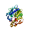


 PDBj
PDBj


 Assembly
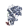
 Mass: 151.126 Da / Num. of mol.: 1 / Source method: obtained synthetically / Formula: C5H5N5O
 Mass: 18.015 Da / Num. of mol.: 258 / Source method: isolated from a natural source / Formula: H2O
 Sample preparation
 / Beamline: BM14 / Wavelength: 0.97 Å
 Processing