 Movie
Movie Controller

[English] 日本語
 Yorodumi
- PDB-3mji: Activation of catalytic cysteine without a base in a Mutant Penic... -
+ Open data

- Basic information
| Entry | Database: PDB / ID: 3mji | ||||||
|---|---|---|---|---|---|---|---|
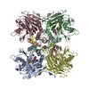
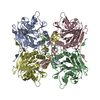
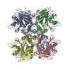
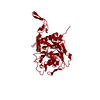
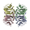
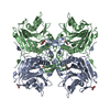
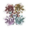
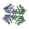
| Title | Activation of catalytic cysteine without a base in a Mutant Penicillin Acylase Precursor | ||||||
 Components Components | Penicillin acylase Penicillin amidase Penicillin amidase | ||||||
 Keywords Keywords | HYDROLASE / ZYMOGEN / PENICILLIN / AUTOPROTEOLYSIS / ANTIBIOTIC RESISTANCE / CATALYSIS / PENICILLIN V ACYLASE / AMIDASE | ||||||
| Function / homology |  Function and homology informationpenicillin amidase / penicillin amidase activity / response to antibiotic Function and homology informationpenicillin amidase / penicillin amidase activity / response to antibioticSimilarity search - Function | ||||||
| Biological species |  Bacillus sphaericus (bacteria) Bacillus sphaericus (bacteria) | ||||||
| Method | X-RAY DIFFRACTION / SYNCHROTRON / MOLECULAR REPLACEMENT / Resolution: 2.5 Å | ||||||
 Authors Authors | Pathak, M.C. / Suresh, C.G. / Dodson, G.G. / Murshudov, G.N. | ||||||
 Citation Citation | Journal: To be Published Title: Activation of catalytic cysteine without a base in a Mutant Penicillin Acylase Precursor Authors: Pathak, M.C. / Suresh, C.G. / Dodson, G.G. / Murshudov, G.N. #1: Journal: Acta Crystallogr.,Sect.F / Year: 2005 Title: Cloning, preparation and preliminary crystallographic studies of Penicillin V Acylase autoproteolytic processing mutants Authors: Chandra, P.M. / Brannigan, J.A. / Prabhune, A. / Pundle, A. / Turkenburg, J.P. / Dodson, G.G. / Suresh, C.G. | ||||||
| History |
|
- Structure visualization
| Structure viewer | Molecule: MolmilJmol/JSmol |
|---|
- Downloads & links
-Download
| PDBx/mmCIF format | 3mji.cif.gz | 263.4 KB | Display | PDBx/mmCIF format |
|---|---|---|---|---|
| PDB format | pdb3mji.ent.gz | 214.8 KB | Display | PDB format |
| PDBx/mmJSON format | 3mji.json.gz | Tree view | PDBx/mmJSON format | |
| Others |  Other downloads Other downloads |
-Validation report
| Arichive directory | https://data.pdbj.org/pub/pdb/validation_reports/mj/3mjiftp://data.pdbj.org/pub/pdb/validation_reports/mj/3mji | HTTPS FTP |
|---|
-Related structure data
| Related structure data |  3mik |
|---|---|
| Similar structure data |


-Links
 PDBj
PDBj


- Assembly
| Deposited unit | 
| ||||||||
|---|---|---|---|---|---|---|---|---|---|
| 1 |
| ||||||||
| Unit cell |
|
-Components
| #1: Protein | Penicillin amidase / Penicillin V amidase / PVA Mass: 37530.535 Da / Num. of mol.: 4 Source method: isolated from a genetically manipulated source Source: (gene. exp.) Bacillus sphaericus (bacteria) / Strain: NCIM 2478 / Production host: Escherichia coli (E. coli) / References: UniProt: P12256, penicillin amidase#2: Water | ChemComp-HOH / | Water Mass: 18.015 Da / Num. of mol.: 319 / Source method: isolated from a natural source / Formula: H2O Mass: 18.015 Da / Num. of mol.: 319 / Source method: isolated from a natural source / Formula: H2O |
|---|
-Experimental details
-Experiment
| Experiment | Method: X-RAY DIFFRACTION / Number of used crystals: 1 |
|---|
- Sample preparation
| Crystal | Density Matthews: 3.08 Å3/Da / Density % sol: 60.06 % Description: THE STRUCTURE FACTOR FILE CONTAINS FRIEDEL PAIRS |
|---|---|
| Crystal grow | Method: vapor diffusion, hanging drop / pH: 6.4 / Details: pH 6.4, VAPOR DIFFUSION, HANGING DROP |
-Data collection
| Diffraction | Mean temperature: 100 K | |||||||||||||||
|---|---|---|---|---|---|---|---|---|---|---|---|---|---|---|---|---|
| Diffraction source | Source: SYNCHROTRON / Site: SRS  / Beamline: PX14.2 / Wavelength: 0.978 Å / Beamline: PX14.2 / Wavelength: 0.978 Å | |||||||||||||||
| Detector | Detector: CCD | |||||||||||||||
| Radiation | Protocol: SINGLE WAVELENGTH / Monochromatic (M) / Laue (L): M / Scattering type: x-ray | |||||||||||||||
| Radiation wavelength | Wavelength: 0.978 Å / Relative weight: 1 | |||||||||||||||
| Reflection twin |
| |||||||||||||||
| Reflection | Resolution: 2.5→50 Å / Num. obs: 62997 / % possible obs: 99.9 % / Rsym value: 0.086 |
- Processing
| Software |
| ||||||||||||||||||||||||||||||||||||||||||||||||||||||||||||||||||||||||||||||||||||||||||||||||||||||||||||||||||||||||||||||||||||||||||||||||||||||||||||||||||||||||||
|---|---|---|---|---|---|---|---|---|---|---|---|---|---|---|---|---|---|---|---|---|---|---|---|---|---|---|---|---|---|---|---|---|---|---|---|---|---|---|---|---|---|---|---|---|---|---|---|---|---|---|---|---|---|---|---|---|---|---|---|---|---|---|---|---|---|---|---|---|---|---|---|---|---|---|---|---|---|---|---|---|---|---|---|---|---|---|---|---|---|---|---|---|---|---|---|---|---|---|---|---|---|---|---|---|---|---|---|---|---|---|---|---|---|---|---|---|---|---|---|---|---|---|---|---|---|---|---|---|---|---|---|---|---|---|---|---|---|---|---|---|---|---|---|---|---|---|---|---|---|---|---|---|---|---|---|---|---|---|---|---|---|---|---|---|---|---|---|---|---|---|---|
| Refinement | Method to determine structure: MOLECULAR REPLACEMENT / Resolution: 2.5→19.95 Å / Cor.coef. Fo:Fc: 0.949 / Cor.coef. Fo:Fc free: 0.912 / SU B: 6.938 / SU ML: 0.151 / Cross valid method: THROUGHOUT / ESU R: 0.072 / ESU R Free: 0.048 / Stereochemistry target values: MAXIMUM LIKELIHOOD / Details: HYDROGENS HAVE BEEN USED IF PRESENT IN THE INPUT
| ||||||||||||||||||||||||||||||||||||||||||||||||||||||||||||||||||||||||||||||||||||||||||||||||||||||||||||||||||||||||||||||||||||||||||||||||||||||||||||||||||||||||||
| Solvent computation | Ion probe radii: 0.8 Å / Shrinkage radii: 0.8 Å / VDW probe radii: 1.2 Å / Solvent model: MASK | ||||||||||||||||||||||||||||||||||||||||||||||||||||||||||||||||||||||||||||||||||||||||||||||||||||||||||||||||||||||||||||||||||||||||||||||||||||||||||||||||||||||||||
| Displacement parameters | Biso mean: 27.443 Å2
| ||||||||||||||||||||||||||||||||||||||||||||||||||||||||||||||||||||||||||||||||||||||||||||||||||||||||||||||||||||||||||||||||||||||||||||||||||||||||||||||||||||||||||
| Refinement step | Cycle: LAST / Resolution: 2.5→19.95 Å
| ||||||||||||||||||||||||||||||||||||||||||||||||||||||||||||||||||||||||||||||||||||||||||||||||||||||||||||||||||||||||||||||||||||||||||||||||||||||||||||||||||||||||||
| Refine LS restraints |
| ||||||||||||||||||||||||||||||||||||||||||||||||||||||||||||||||||||||||||||||||||||||||||||||||||||||||||||||||||||||||||||||||||||||||||||||||||||||||||||||||||||||||||
| LS refinement shell | Resolution: 2.5→2.557 Å / Total num. of bins used: 20
|
