 Movie
Movie Controller

[English] 日本語
 Yorodumi
- PDB-3mbl: Crystal Structure of the human mitogen-activated protein kinase k... -
+ Open data

- Basic information
| Entry | Database: PDB / ID: 3mbl | ||||||
|---|---|---|---|---|---|---|---|
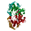
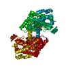
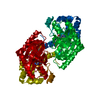
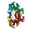
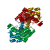
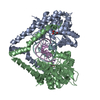
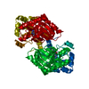
| Title | Crystal Structure of the human mitogen-activated protein kinase kinase 1 (MEK 1) in complex with ligand and MgADP | ||||||
 Components Components | Dual specificity mitogen-activated protein kinase kinase 1 | ||||||
 Keywords Keywords |  TRANSFERASE / Kinase inhibitor TRANSFERASE / Kinase inhibitor | ||||||
| Function / homology |  Function and homology information Function and homology informationepithelial cell proliferation involved in lung morphogenesis / positive regulation of endodermal cell differentiation / placenta blood vessel development / regulation of axon regeneration / mitogen-activated protein kinase kinase / type B pancreatic cell proliferation / labyrinthine layer development / MAP-kinase scaffold activity / cerebellar cortex formation / Signaling by MAP2K mutants ...epithelial cell proliferation involved in lung morphogenesis / positive regulation of endodermal cell differentiation / placenta blood vessel development / regulation of axon regeneration / mitogen-activated protein kinase kinase / type B pancreatic cell proliferation / labyrinthine layer development / MAP-kinase scaffold activity / cerebellar cortex formation / Signaling by MAP2K mutants / regulation of Golgi inheritance / trachea formation / regulation of early endosome to late endosome transport / Negative feedback regulation of MAPK pathway / regulation of stress-activated MAPK cascade / positive regulation of axonogenesis / Frs2-mediated activation / protein kinase activator activity / ERBB2-ERBB3 signaling pathway / face development / endodermal cell differentiation / MAPK3 (ERK1) activation / Bergmann glial cell differentiation / MAP kinase kinase activity / thyroid gland development / Uptake and function of anthrax toxins / Schwann cell development / keratinocyte differentiation / ERK1 and ERK2 cascade / myelination / protein serine/threonine/tyrosine kinase activity / protein serine/threonine kinase activator activity / MAP3K8 (TPL2)-dependent MAPK1/3 activation / insulin-like growth factor receptor signaling pathway / thymus development / Signal transduction by L1 / cell motility / RAF activation / Signaling by high-kinase activity BRAF mutants / MAP2K and MAPK activation / positive regulation of protein serine/threonine kinase activity / neuron differentiation / Signaling by RAF1 mutants / Signaling by moderate kinase activity BRAF mutants / Paradoxical activation of RAF signaling by kinase inactive BRAF / Signaling downstream of RAS mutants / chemotaxis / MAPK cascade / cellular senescence / Signaling by BRAF and RAF1 fusions / late endosome / heart development / scaffold protein binding / protein tyrosine kinase activity / positive regulation of ERK1 and ERK2 cascade / early endosome / protein kinase activity / negative regulation of cell population proliferation / protein phosphorylation / focal adhesion / protein serine kinase activity / centrosome / protein serine/threonine kinase activity / positive regulation of gene expression / positive regulation of DNA-templated transcription / Golgi apparatus / endoplasmic reticulum / signal transduction / mitochondrion / ATP binding / nucleus / plasma membrane / cytosolSimilarity search - Function | ||||||
| Biological species |  Homo sapiens (human) Homo sapiens (human) | ||||||
| Method | X-RAY DIFFRACTION / SYNCHROTRON / MOLECULAR REPLACEMENT / Resolution: 2.6 Å | ||||||
 Authors Authors | Dougan, D.R. / Mol, C.D. | ||||||
 Citation Citation | Journal: Bioorg.Med.Chem.Lett. / Year: 2010 Title: Structure-based design and synthesis of pyrrole derivatives as MEK inhibitors. Authors: Wallace, M.B. / Adams, M.E. / Kanouni, T. / Mol, C.D. / Dougan, D.R. / Feher, V.A. / O'Connell, S.M. / Shi, L. / Halkowycz, P. / Dong, Q. | ||||||
| History |
|
- Structure visualization
| Structure viewer | Molecule: MolmilJmol/JSmol |
|---|
- Downloads & links
-Download
| PDBx/mmCIF format | 3mbl.cif.gz | 135.8 KB | Display | PDBx/mmCIF format |
|---|---|---|---|---|
| PDB format | pdb3mbl.ent.gz | 106.4 KB | Display | PDB format |
| PDBx/mmJSON format | 3mbl.json.gz | Tree view | PDBx/mmJSON format | |
| Others |  Other downloads Other downloads |
-Validation report
| Arichive directory | https://data.pdbj.org/pub/pdb/validation_reports/mb/3mblftp://data.pdbj.org/pub/pdb/validation_reports/mb/3mbl | HTTPS FTP |
|---|
-Related structure data
| Similar structure data |
|---|


-Links
 PDBj
PDBj


- Assembly
| Deposited unit | 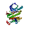
| ||||||||
|---|---|---|---|---|---|---|---|---|---|
| 1 | 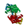
| ||||||||
| 2 |
| ||||||||
| Unit cell |
|
-Components
| #1: Protein | Mass: 36640.219 Da / Num. of mol.: 1 / Fragment: Kinase Domain Source method: isolated from a genetically manipulated source Source: (gene. exp.) Homo sapiens (human) / Gene: MAP2K1, MEK 1, MEK1, PRKMK1 / Plasmid: pET21 / Production host:  Escherichia coli (E. coli) / Strain (production host): BL21(DE3) Escherichia coli (E. coli) / Strain (production host): BL21(DE3)References: UniProt: Q02750, mitogen-activated protein kinase kinase |
|---|---|
| #2: Chemical | ChemComp-ADP / Adenosine diphosphate  Mass: 427.201 Da / Num. of mol.: 1 / Source method: obtained synthetically / Formula: C10H15N5O10P2 / Comment: ADP, energy-carrying molecule*YM Mass: 427.201 Da / Num. of mol.: 1 / Source method: obtained synthetically / Formula: C10H15N5O10P2 / Comment: ADP, energy-carrying molecule*YM |
| #3: Chemical | ChemComp-MG /   Mass: 24.305 Da / Num. of mol.: 1 / Source method: obtained synthetically / Formula: Mg Mass: 24.305 Da / Num. of mol.: 1 / Source method: obtained synthetically / Formula: Mg |
| #4: Chemical | ChemComp-LSG /   Mass: 461.227 Da / Num. of mol.: 1 / Source method: obtained synthetically / Formula: C16H17FIN3O4 Mass: 461.227 Da / Num. of mol.: 1 / Source method: obtained synthetically / Formula: C16H17FIN3O4 |
| #5: Water | ChemComp-HOH / Water Mass: 18.015 Da / Num. of mol.: 90 / Source method: isolated from a natural source / Formula: H2O Mass: 18.015 Da / Num. of mol.: 90 / Source method: isolated from a natural source / Formula: H2O |
-Experimental details
-Experiment
| Experiment | Method: X-RAY DIFFRACTION / Number of used crystals: 1 |
|---|
- Sample preparation
| Crystal | Density Matthews: 3.44 Å3/Da / Density % sol: 64.24 % |
|---|---|
| Crystal grow | Temperature: 298 K / Method: vapor diffusion, sitting drop / Details: VAPOR DIFFUSION, SITTING DROP, temperature 298K |
-Data collection
| Diffraction | Mean temperature: 100 K |
|---|---|
| Diffraction source | Source: SYNCHROTRON / Site: ALS  / Beamline: 5.0.3 / Wavelength: 1 Å / Beamline: 5.0.3 / Wavelength: 1 Å |
| Detector | Type: ADSC QUANTUM 315r / Detector: CCD / Date: Sep 2, 2006 |
| Radiation | Protocol: SINGLE WAVELENGTH / Monochromatic (M) / Laue (L): M / Scattering type: x-ray |
| Radiation wavelength | Wavelength: 1 Å / Relative weight: 1 |
| Reflection | Resolution: 2.6→50 Å / Num. obs: 15253 / % possible obs: 98.5 % / Redundancy: 2.5 % / Rmerge(I) obs: 0.055 / Net I/σ(I): 24.4 |
| Reflection shell | Resolution: 2.6→2.68 Å / Rmerge(I) obs: 0.524 / Mean I/σ(I) obs: 2.4 |
- Processing
| Software |
| ||||||||||||||||||||||||||||||||||||||||||||||||||||||||||||||||||||||||||||||||||||||||||||||||||||||||||||||||||||||||||||||||||||||||||||||||||||||||||||||||||||||||||
|---|---|---|---|---|---|---|---|---|---|---|---|---|---|---|---|---|---|---|---|---|---|---|---|---|---|---|---|---|---|---|---|---|---|---|---|---|---|---|---|---|---|---|---|---|---|---|---|---|---|---|---|---|---|---|---|---|---|---|---|---|---|---|---|---|---|---|---|---|---|---|---|---|---|---|---|---|---|---|---|---|---|---|---|---|---|---|---|---|---|---|---|---|---|---|---|---|---|---|---|---|---|---|---|---|---|---|---|---|---|---|---|---|---|---|---|---|---|---|---|---|---|---|---|---|---|---|---|---|---|---|---|---|---|---|---|---|---|---|---|---|---|---|---|---|---|---|---|---|---|---|---|---|---|---|---|---|---|---|---|---|---|---|---|---|---|---|---|---|---|---|---|
| Refinement | Method to determine structure: MOLECULAR REPLACEMENT / Resolution: 2.6→20 Å / Cor.coef. Fo:Fc: 0.971 / Cor.coef. Fo:Fc free: 0.958 / SU B: 17.217 / SU ML: 0.188 / Cross valid method: THROUGHOUT / ESU R: 0.317 / ESU R Free: 0.234 / Stereochemistry target values: MAXIMUM LIKELIHOOD / Details: HYDROGENS HAVE BEEN ADDED IN THE RIDING POSITIONS
| ||||||||||||||||||||||||||||||||||||||||||||||||||||||||||||||||||||||||||||||||||||||||||||||||||||||||||||||||||||||||||||||||||||||||||||||||||||||||||||||||||||||||||
| Solvent computation | Ion probe radii: 0.8 Å / Shrinkage radii: 0.8 Å / VDW probe radii: 1.4 Å / Solvent model: MASK | ||||||||||||||||||||||||||||||||||||||||||||||||||||||||||||||||||||||||||||||||||||||||||||||||||||||||||||||||||||||||||||||||||||||||||||||||||||||||||||||||||||||||||
| Displacement parameters | Biso mean: 78.531 Å2
| ||||||||||||||||||||||||||||||||||||||||||||||||||||||||||||||||||||||||||||||||||||||||||||||||||||||||||||||||||||||||||||||||||||||||||||||||||||||||||||||||||||||||||
| Refinement step | Cycle: LAST / Resolution: 2.6→20 Å
| ||||||||||||||||||||||||||||||||||||||||||||||||||||||||||||||||||||||||||||||||||||||||||||||||||||||||||||||||||||||||||||||||||||||||||||||||||||||||||||||||||||||||||
| Refine LS restraints |
| ||||||||||||||||||||||||||||||||||||||||||||||||||||||||||||||||||||||||||||||||||||||||||||||||||||||||||||||||||||||||||||||||||||||||||||||||||||||||||||||||||||||||||
| LS refinement shell | Resolution: 2.6→2.667 Å / Total num. of bins used: 20
| ||||||||||||||||||||||||||||||||||||||||||||||||||||||||||||||||||||||||||||||||||||||||||||||||||||||||||||||||||||||||||||||||||||||||||||||||||||||||||||||||||||||||||
| Refinement TLS params. | Method: refined / Refine-ID: X-RAY DIFFRACTION
| ||||||||||||||||||||||||||||||||||||||||||||||||||||||||||||||||||||||||||||||||||||||||||||||||||||||||||||||||||||||||||||||||||||||||||||||||||||||||||||||||||||||||||
| Refinement TLS group |
|
