 Movie
Movie Controller

[English] 日本語
 Yorodumi
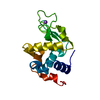
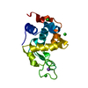
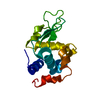
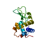
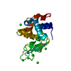
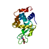
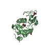
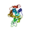
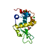
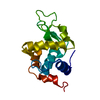
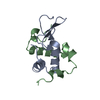
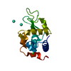
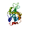
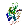
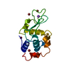
- PDB-3m3u: Effect of temperature on tryptophan fluorescence in lysozyme crystals -
+ Open data

- Basic information
| Entry | Database: PDB / ID: 3m3u | ||||||
|---|---|---|---|---|---|---|---|
| Title | Effect of temperature on tryptophan fluorescence in lysozyme crystals | ||||||
 Components Components | Lysozyme C | ||||||
 Keywords Keywords |  HYDROLASE / Hen egg white lysozyme / Tryptophan fluorescence / Allergen / Antimicrobial / Bacteriolytic enzyme / Disulfide bond / Glycosidase HYDROLASE / Hen egg white lysozyme / Tryptophan fluorescence / Allergen / Antimicrobial / Bacteriolytic enzyme / Disulfide bond / Glycosidase | ||||||
| Function / homology |  Function and homology informationAntimicrobial peptides / Neutrophil degranulation / beta-N-acetylglucosaminidase activity / cell wall macromolecule catabolic process / lysozyme / lysozyme activity / killing of cells of another organism / defense response to Gram-negative bacterium / defense response to Gram-positive bacterium / defense response to bacterium ...Antimicrobial peptides / Neutrophil degranulation / beta-N-acetylglucosaminidase activity / cell wall macromolecule catabolic process / lysozyme / lysozyme activity / killing of cells of another organism / defense response to Gram-negative bacterium / defense response to Gram-positive bacterium / defense response to bacterium / endoplasmic reticulum / extracellular space / identical protein binding / cytoplasm Function and homology informationAntimicrobial peptides / Neutrophil degranulation / beta-N-acetylglucosaminidase activity / cell wall macromolecule catabolic process / lysozyme / lysozyme activity / killing of cells of another organism / defense response to Gram-negative bacterium / defense response to Gram-positive bacterium / defense response to bacterium ...Antimicrobial peptides / Neutrophil degranulation / beta-N-acetylglucosaminidase activity / cell wall macromolecule catabolic process / lysozyme / lysozyme activity / killing of cells of another organism / defense response to Gram-negative bacterium / defense response to Gram-positive bacterium / defense response to bacterium / endoplasmic reticulum / extracellular space / identical protein binding / cytoplasmSimilarity search - Function | ||||||
| Biological species |  Gallus gallus (chicken) Gallus gallus (chicken) | ||||||
| Method | X-RAY DIFFRACTION / MOLECULAR REPLACEMENT / Resolution: 2.35 Å | ||||||
 Authors Authors | Varshney, N.K. / Sitaramam, V. | ||||||
 Citation Citation | Journal: TO BE PUBLISHED Title: Effect of temperature on tryptophan fluorescence in lysozyme crystals Authors: Varshney, N.K. / Sitaramam, V. #1: Journal: J.Biol.Chem. / Year: 1990Title: Crystal structure of low humidity tetragonal lysozyme at 2.1-A resolution. Variability in hydration shell and its structural consequences. Authors: Kodandapani, R. / Suresh, C.G. / Vijayan, M. | ||||||
| History |
|
- Structure visualization
| Structure viewer | Molecule: MolmilJmol/JSmol |
|---|
- Downloads & links
-Download
| PDBx/mmCIF format | 3m3u.cif.gz | 40.1 KB | Display | PDBx/mmCIF format |
|---|---|---|---|---|
| PDB format | pdb3m3u.ent.gz | 27.1 KB | Display | PDB format |
| PDBx/mmJSON format | 3m3u.json.gz | Tree view | PDBx/mmJSON format | |
| Others |  Other downloads Other downloads |
-Validation report
| Arichive directory | https://data.pdbj.org/pub/pdb/validation_reports/m3/3m3uftp://data.pdbj.org/pub/pdb/validation_reports/m3/3m3u | HTTPS FTP |
|---|
-Related structure data
| Related structure data | 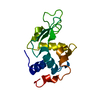 1bvxS S: Starting model for refinement |
|---|---|
| Similar structure data |

-Links
 PDBj
PDBj


- Assembly
| Deposited unit | 
| ||||||||
|---|---|---|---|---|---|---|---|---|---|
| 1 |
| ||||||||
| Unit cell |
|
-Components
| #1: Protein | Mass: 14331.160 Da / Num. of mol.: 1 / Source method: isolated from a natural source / Source: (natural) Gallus gallus (chicken) / Tissue: egg white / References: UniProt: P00698, lysozyme |
|---|---|
| #2: Chemical | ChemComp-NA /   Mass: 22.990 Da / Num. of mol.: 1 / Source method: obtained synthetically / Formula: Na Mass: 22.990 Da / Num. of mol.: 1 / Source method: obtained synthetically / Formula: Na |
| #3: Chemical | ChemComp-CL / Chloride  Mass: 35.453 Da / Num. of mol.: 1 / Source method: obtained synthetically / Formula: Cl Mass: 35.453 Da / Num. of mol.: 1 / Source method: obtained synthetically / Formula: Cl |
| #4: Water | ChemComp-HOH / Water Mass: 18.015 Da / Num. of mol.: 64 / Source method: isolated from a natural source / Formula: H2O Mass: 18.015 Da / Num. of mol.: 64 / Source method: isolated from a natural source / Formula: H2O |
-Experimental details
-Experiment
| Experiment | Method: X-RAY DIFFRACTION / Number of used crystals: 1 |
|---|
- Sample preparation
| Crystal | Density Matthews: 2.08 Å3/Da / Density % sol: 40.85 % |
|---|---|
| Crystal grow | Temperature: 295 K / Method: vapor diffusion, hanging drop / pH: 4.8 Details: 0.1M Sodium Acetate, 7% NaCl, 25% Ethylene glycol, pH 4.8, VAPOR DIFFUSION, HANGING DROP, temperature 295K |
-Data collection
| Diffraction | Mean temperature: 293 K |
|---|---|
| Diffraction source | Source: ROTATING ANODE / Type: RIGAKU / Wavelength: 1.514 Å |
| Detector | Type: RIGAKU RAXIS IV / Detector: IMAGE PLATE / Date: Jul 16, 2005 / Details: Mirrors |
| Radiation | Monochromator: Osmic mirror / Protocol: SINGLE WAVELENGTH / Monochromatic (M) / Laue (L): M / Scattering type: x-ray |
| Radiation wavelength | Wavelength: 1.514 Å / Relative weight: 1 |
| Reflection | Resolution: 2.35→19.77 Å / Num. all: 5388 / Num. obs: 5384 / % possible obs: 99.93 % / Observed criterion σ(F): 1 / Redundancy: 12.86 % / Biso Wilson estimate: 35.74 Å2 / Rmerge(I) obs: 0.065 / Net I/σ(I): 11.26 |
| Reflection shell | Resolution: 2.349→2.409 Å / Redundancy: 10.43 % / Rmerge(I) obs: 0.163 / Mean I/σ(I) obs: 5.211 / Num. unique all: 363 / % possible all: 99.21 |
- Processing
| Software |
| |||||||||||||||||||||||||||||||||||||||||||||||||||||||||||||||||
|---|---|---|---|---|---|---|---|---|---|---|---|---|---|---|---|---|---|---|---|---|---|---|---|---|---|---|---|---|---|---|---|---|---|---|---|---|---|---|---|---|---|---|---|---|---|---|---|---|---|---|---|---|---|---|---|---|---|---|---|---|---|---|---|---|---|---|
| Refinement | Method to determine structure: MOLECULAR REPLACEMENT Starting model: 1BVX Resolution: 2.35→19.77 Å / Cor.coef. Fo:Fc: 0.95 / Cor.coef. Fo:Fc free: 0.938 / Cross valid method: THROUGHOUT / σ(F): 1 / ESU R: 0.424 / ESU R Free: 0.215 / Stereochemistry target values: MAXIMUM LIKELIHOOD / Details: HYDROGENS HAVE BEEN ADDED IN THE RIDING POSITIONS
| |||||||||||||||||||||||||||||||||||||||||||||||||||||||||||||||||
| Solvent computation | Ion probe radii: 0.8 Å / Shrinkage radii: 0.8 Å / VDW probe radii: 1.4 Å / Solvent model: MASK | |||||||||||||||||||||||||||||||||||||||||||||||||||||||||||||||||
| Displacement parameters | Biso mean: 21.524 Å2
| |||||||||||||||||||||||||||||||||||||||||||||||||||||||||||||||||
| Refinement step | Cycle: LAST / Resolution: 2.35→19.77 Å
| |||||||||||||||||||||||||||||||||||||||||||||||||||||||||||||||||
| Refine LS restraints |
| |||||||||||||||||||||||||||||||||||||||||||||||||||||||||||||||||
| LS refinement shell | Resolution: 2.349→2.409 Å / Total num. of bins used: 20
|
