 Movie
Movie Controller

[English] 日本語
 Yorodumi
- PDB-3lra: Structural Basis for Assembling a Human Tripartite Complex Dlg1-M... -
+ Open data

- Basic information
| Entry | Database: PDB / ID: 3lra | ||||||
|---|---|---|---|---|---|---|---|
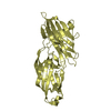
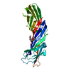
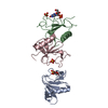
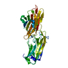
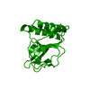
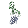
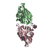
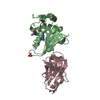
| Title | Structural Basis for Assembling a Human Tripartite Complex Dlg1-MPP7-Mals3 | ||||||
 Components Components | Disks large homolog 1, MAGUK p55 subfamily member 7, Protein lin-7 homolog C | ||||||
 Keywords Keywords |  MEMBRANE PROTEIN / Tripartite Complex / L27 tetramer / Cell junction / Cell membrane / Endoplasmic reticulum / Host-virus interaction / Postsynaptic cell membrane / SH3 domain / Synapse / Tight junction / Exocytosis / Protein transport / Synaptosome / Transport MEMBRANE PROTEIN / Tripartite Complex / L27 tetramer / Cell junction / Cell membrane / Endoplasmic reticulum / Host-virus interaction / Postsynaptic cell membrane / SH3 domain / Synapse / Tight junction / Exocytosis / Protein transport / Synaptosome / Transport | ||||||
| Function / homology |  Function and homology information Function and homology informationprotein localization to basolateral plasma membrane / regulation of voltage-gated potassium channel activity involved in ventricular cardiac muscle cell action potential repolarization / regulation of protein localization to synapse / regulation of potassium ion import / L27 domain binding / regulation of potassium ion export across plasma membrane / MPP7-DLG1-LIN7 complex / morphogenesis of an epithelial sheet / membrane raft organization / hard palate development ...protein localization to basolateral plasma membrane / regulation of voltage-gated potassium channel activity involved in ventricular cardiac muscle cell action potential repolarization / regulation of protein localization to synapse / regulation of potassium ion import / L27 domain binding / regulation of potassium ion export across plasma membrane / MPP7-DLG1-LIN7 complex / morphogenesis of an epithelial sheet / membrane raft organization / hard palate development / maintenance of epithelial cell apical/basal polarity / establishment of centrosome localization / cortical microtubule organization / negative regulation of p38MAPK cascade / guanylate kinase activity / NrCAM interactions / regulation of sodium ion transmembrane transport / Dopamine Neurotransmitter Release Cycle / embryonic skeletal system morphogenesis / astral microtubule organization / structural constituent of postsynaptic density / lateral loop / reproductive structure development / myelin sheath abaxonal region / immunological synapse formation / peristalsis / Synaptic adhesion-like molecules / protein localization to adherens junction / cell projection membrane / smooth muscle tissue development / bicellular tight junction assembly / positive regulation of potassium ion transport / neurotransmitter secretion / regulation of ventricular cardiac muscle cell action potential / amyloid precursor protein metabolic process / node of Ranvier / Trafficking of AMPA receptors / establishment or maintenance of epithelial cell apical/basal polarity / protein-containing complex localization / Assembly and cell surface presentation of NMDA receptors / Neurexins and neuroligins / endothelial cell proliferation / neurotransmitter receptor localization to postsynaptic specialization membrane / lens development in camera-type eye / regulation of myelination / Activation of Ca-permeable Kainate Receptor / cortical actin cytoskeleton organization / establishment or maintenance of cell polarity / branching involved in ureteric bud morphogenesis / negative regulation of G1/S transition of mitotic cell cycle / establishment of cell polarity / positive regulation of actin filament polymerization / receptor clustering / Negative regulation of NMDA receptor-mediated neuronal transmission / Unblocking of NMDA receptors, glutamate binding and activation / RHOJ GTPase cycle / RHOQ GTPase cycle / exocytosis / phosphoprotein phosphatase activity / Long-term potentiation / immunological synapse / RHOG GTPase cycle / basement membrane / negative regulation of phosphatidylinositol 3-kinase/protein kinase B signal transduction / bicellular tight junction / intercalated disc / potassium channel regulator activity / lateral plasma membrane / RAC3 GTPase cycle / RAC2 GTPase cycle / phosphatase binding / signaling adaptor activity / T cell proliferation / negative regulation of T cell proliferation / RAC1 GTPase cycle / cytoskeletal protein binding / actin filament polymerization / Ras activation upon Ca2+ influx through NMDA receptor / regulation of membrane potential / phosphatidylinositol 3-kinase/protein kinase B signal transduction / synaptic membrane / actin filament organization / PDZ domain binding / protein localization to plasma membrane / positive regulation of protein-containing complex assembly / postsynaptic density membrane / positive regulation of protein localization to plasma membrane / adherens junction / sarcolemma / neuromuscular junction / cytoplasmic side of plasma membrane / negative regulation of ERK1 and ERK2 cascade / cell-cell adhesion / kinase binding / negative regulation of epithelial cell proliferation / cell-cell junction / protein transport / presynapse / cell junction / cell cortexSimilarity search - Function | ||||||
| Biological species |  Homo sapiens (human) Homo sapiens (human) | ||||||
| Method | X-RAY DIFFRACTION / SYNCHROTRON / SAD / Resolution: 2.95 Å | ||||||
 Authors Authors | Yang, X. / Xie, X. / Shen, Y. / Long, J. | ||||||
 Citation Citation | Journal: Faseb J. / Year: 2010 Title: Structural basis for tandem L27 domain-mediated polymerization Authors: Yang, X. / Xie, X. / Chen, L. / Zhou, H. / Wang, Z. / Zhao, W. / Tian, R. / Zhang, R. / Tian, C. / Long, J. / Shen, Y. | ||||||
| History |
|
- Structure visualization
| Structure viewer | Molecule: MolmilJmol/JSmol |
|---|
- Downloads & links
-Download
| PDBx/mmCIF format | 3lra.cif.gz | 63.5 KB | Display | PDBx/mmCIF format |
|---|---|---|---|---|
| PDB format | pdb3lra.ent.gz | 47.5 KB | Display | PDB format |
| PDBx/mmJSON format | 3lra.json.gz | Tree view | PDBx/mmJSON format | |
| Others |  Other downloads Other downloads |
-Validation report
| Arichive directory | https://data.pdbj.org/pub/pdb/validation_reports/lr/3lraftp://data.pdbj.org/pub/pdb/validation_reports/lr/3lra | HTTPS FTP |
|---|
-Related structure data
| Similar structure data |
|---|


-Links
 PDBj
PDBj


- Assembly
| Deposited unit | 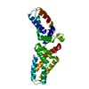
| ||||||||
|---|---|---|---|---|---|---|---|---|---|
| 1 |
| ||||||||
| Unit cell |
|
-Components
| #1: Protein | Mass: 29164.166 Da / Num. of mol.: 1 / Fragment: L27 domain, L27 1/2 domain Source method: isolated from a genetically manipulated source Details: Structural Basis for Assembling a Tripartite Complex Dlg1-MPP7-Mals3 Source: (gene. exp.) Homo sapiens (human) / Gene: L27 domains of Dlg1, MPP7 and Mals3Plasmid details: in-house-modified version of the pET32a vector (Novagen), in which the S-tag an d the thrombin recognition site were replaced by a sequence encoding a TEV prote ase cleavage site ...Plasmid details: in-house-modified version of the pET32a vector (Novagen), in which the S-tag an d the thrombin recognition site were replaced by a sequence encoding a TEV prote ase cleavage site (Glu-Asn-Leu-Tyr-Phe-Gln-Ser) Plasmid: pET32a vector (Novagen) / Production host:  Escherichia coli (E. coli) / Strain (production host): BL21(DE3) Escherichia coli (E. coli) / Strain (production host): BL21(DE3)References: UniProt: Q12959, UniProt: Q5T2T1, UniProt: Q9NUP9 |
|---|---|
| #2: Water | ChemComp-HOH / Water Mass: 18.015 Da / Num. of mol.: 41 / Source method: isolated from a natural source / Formula: H2O Mass: 18.015 Da / Num. of mol.: 41 / Source method: isolated from a natural source / Formula: H2O |
| Compound details | THE L27 DOMAINS OF HUMAN DLG1 (TERMED AS L27HDLG1), HUMAN MPP7 (TERMED AS L27N AND L27C) AND HUMAN ...THE L27 DOMAINS OF HUMAN DLG1 (TERMED AS L27HDLG1), HUMAN MPP7 (TERMED AS L27N AND L27C) AND HUMAN MALS3 (TERMED AS L27HMALS3) WERE COVALENTLY |
| Sequence details | THE INTERNAL SEQUENCE LEVLFQGP ARE LINKER. |
-Experimental details
-Experiment
| Experiment | Method: X-RAY DIFFRACTION / Number of used crystals: 2 |
|---|
- Sample preparation
| Crystal | Density Matthews: 3.42 Å3/Da / Density % sol: 64.04 % |
|---|---|
| Crystal grow | Temperature: 293 K / Method: vapor diffusion, sitting drop / pH: 7 Details: 10% PEG 8000, 0.2M MgCl2, 0.1M Guanidine HCl, 0.1M Tris-HCl, pH7.0, VAPOR DIFFUSION, SITTING DROP, temperature 293K |
-Data collection
| Diffraction |
| ||||||||||||||||||
|---|---|---|---|---|---|---|---|---|---|---|---|---|---|---|---|---|---|---|---|
| Diffraction source |
| ||||||||||||||||||
| Detector |
| ||||||||||||||||||
| Radiation |
| ||||||||||||||||||
| Radiation wavelength |
| ||||||||||||||||||
| Reflection | Resolution: 2.95→38.3 Å / Num. all: 8504 / Num. obs: 8045 / % possible obs: 95 % / Observed criterion σ(F): 5 / Observed criterion σ(I): 2 / Redundancy: 14.8 % / Biso Wilson estimate: 1.2 Å2 / Rmerge(I) obs: 0.092 / Rsym value: 0.092 / Net I/σ(I): 25.7 | ||||||||||||||||||
| Reflection shell | Resolution: 2.95→3.06 Å / Redundancy: 4.5 % / Rmerge(I) obs: 0.435 / Mean I/σ(I) obs: 2.1 / Num. unique all: 1047 / Rsym value: 0.435 / % possible all: 77.7 |


- Processing
| Software |
| ||||||||||||||||||||||||||||||||||||
|---|---|---|---|---|---|---|---|---|---|---|---|---|---|---|---|---|---|---|---|---|---|---|---|---|---|---|---|---|---|---|---|---|---|---|---|---|---|
| Refinement | Method to determine structure: SAD / Resolution: 2.95→38.3 Å / Rfactor Rfree error: 0.013 / Data cutoff high absF: 52963.05 / Data cutoff low absF: 0 / Isotropic thermal model: RESTRAINED / Cross valid method: THROUGHOUT / σ(F): 5 / σ(I): 2 / Stereochemistry target values: Engh & Huber / Details: BULK SOLVENT MODEL USED
| ||||||||||||||||||||||||||||||||||||
| Solvent computation | Solvent model: FLAT MODEL / Bsol: 52.039 Å2 / ksol: 0.35 e/Å3 | ||||||||||||||||||||||||||||||||||||
| Displacement parameters | Biso mean: 67.2 Å2
| ||||||||||||||||||||||||||||||||||||
| Refine analyze |
| ||||||||||||||||||||||||||||||||||||
| Refinement step | Cycle: LAST / Resolution: 2.95→38.3 Å
| ||||||||||||||||||||||||||||||||||||
| Refine LS restraints |
| ||||||||||||||||||||||||||||||||||||
| LS refinement shell | Resolution: 2.95→3.06 Å / Rfactor Rfree error: 0.047 / Total num. of bins used: 6
| ||||||||||||||||||||||||||||||||||||
| Xplor file |
|
