 Movie
Movie Controller

[English] 日本語
 Yorodumi
- PDB-3loi: Crystal structures of Cupin superfamily BbDUF985 from Branchiosto... -
+ Open data

- Basic information
| Entry | Database: PDB / ID: 3loi | ||||||
|---|---|---|---|---|---|---|---|
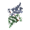
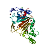
| Title | Crystal structures of Cupin superfamily BbDUF985 from Branchiostoma belcheri tsingtauense in the apo and GDP-bound forms | ||||||
 Components Components | Putative uncharacterized protein | ||||||
 Keywords Keywords | UNKNOWN FUNCTION /  Beta barrel Beta barrel | ||||||
| Function / homology |  Function and homology information Function and homology informationCupin domain of unknown function DUF985 / Uncharacterized protein YML079W-like / Cupin superfamily (DUF985) / RmlC-like cupin domain superfamily / Jelly Rolls / RmlC-like jelly roll fold / Jelly Rolls / Sandwich / Mainly BetaSimilarity search - Domain/homology | ||||||
| Biological species |  | ||||||
| Method | X-RAY DIFFRACTION / SYNCHROTRON / MOLECULAR REPLACEMENT / Resolution: 2.1 Å | ||||||
 Authors Authors | Zou, C.Z. / Du, Y. / He, Y.-X. / Saren, G. / Zhang, X. / Chen, Y. / Zhang, S.-C. | ||||||
 Citation Citation | Journal: Proteins / Year: 2010 Title: Crystal structures of the apo and GDP-bound forms of a cupin-like protein BbDUF985 from Branchiostoma belcheri tsingtauense Authors: Zou, C.Z. / Du, Y. / He, Y.-X. / Saren, G. / Zhang, X. / Chen, Y. / Zhang, S.-C. | ||||||
| History |
|
- Structure visualization
| Structure viewer | Molecule: MolmilJmol/JSmol |
|---|
- Downloads & links
-Download
| PDBx/mmCIF format | 3loi.cif.gz | 49.4 KB | Display | PDBx/mmCIF format |
|---|---|---|---|---|
| PDB format | pdb3loi.ent.gz | 34.4 KB | Display | PDB format |
| PDBx/mmJSON format | 3loi.json.gz | Tree view | PDBx/mmJSON format | |
| Others |  Other downloads Other downloads |
-Validation report
| Arichive directory | https://data.pdbj.org/pub/pdb/validation_reports/lo/3loiftp://data.pdbj.org/pub/pdb/validation_reports/lo/3loi | HTTPS FTP |
|---|
-Related structure data
| Related structure data | 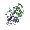 3lzzC 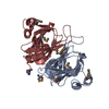 1yudS S: Starting model for refinement C: citing same article ( |
|---|---|
| Similar structure data |


-Links
 PDBj
PDBj- Assembly
| Deposited unit | 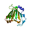
| |||||||||
|---|---|---|---|---|---|---|---|---|---|---|
| 1 | 
| |||||||||
| Unit cell |
| |||||||||
| Components on special symmetry positions |
|
-Components
| #1: Protein | Mass: 19558.209 Da / Num. of mol.: 1 Source method: isolated from a genetically manipulated source Source: (gene. exp.) Plasmid: pET28a / Production host:  Escherichia coli (E. coli) / Strain (production host): BL21(DE3) / References: UniProt: Q6WRH7 Escherichia coli (E. coli) / Strain (production host): BL21(DE3) / References: UniProt: Q6WRH7 |
|---|---|
| #2: Water | ChemComp-HOH / Water Mass: 18.015 Da / Num. of mol.: 173 / Source method: isolated from a natural source / Formula: H2O Mass: 18.015 Da / Num. of mol.: 173 / Source method: isolated from a natural source / Formula: H2O |
-Experimental details
-Experiment
| Experiment | Method: X-RAY DIFFRACTION / Number of used crystals: 1 |
|---|
- Sample preparation
| Crystal | Density Matthews: 4.64 Å3/Da / Density % sol: 73.48 % |
|---|---|
| Crystal grow | Temperature: 289 K / Method: vapor diffusion, hanging drop / pH: 4.6 Details: 2.0M ammonium sulfate, 0.1M sodium acetate trihydate pH 4.6, VAPOR DIFFUSION, HANGING DROP, temperature 289.0K |
-Data collection
| Diffraction | Mean temperature: 100 K |
|---|---|
| Diffraction source | Source: SYNCHROTRON / Site: BSRF  / Beamline: 3W1A / Wavelength: 1 Å / Beamline: 3W1A / Wavelength: 1 Å |
| Detector | Type: MAR CCD 165 mm / Detector: CCD / Date: Jan 10, 2008 |
| Radiation | Protocol: SINGLE WAVELENGTH / Monochromatic (M) / Laue (L): M / Scattering type: x-ray |
| Radiation wavelength | Wavelength: 1 Å / Relative weight: 1 |
| Reflection | Resolution: 2.1→40 Å / Num. obs: 21913 / % possible obs: 99.9 % / Redundancy: 10 % / Biso Wilson estimate: 40.1 Å2 / Rmerge(I) obs: 0.117 / Net I/σ(I): 18.2 |
| Reflection shell | Resolution: 2.1→2.14 Å / Redundancy: 7.3 % / Rmerge(I) obs: 0.577 / Mean I/σ(I) obs: 2.2 / Num. unique all: 1071 / % possible all: 98.5 |
- Processing
| Software |
| |||||||||||||||||||||||||||||||||||||||||||||||||||||||||||||||
|---|---|---|---|---|---|---|---|---|---|---|---|---|---|---|---|---|---|---|---|---|---|---|---|---|---|---|---|---|---|---|---|---|---|---|---|---|---|---|---|---|---|---|---|---|---|---|---|---|---|---|---|---|---|---|---|---|---|---|---|---|---|---|---|---|
| Refinement | Method to determine structure: MOLECULAR REPLACEMENT Starting model: 1YUD Resolution: 2.1→33.396 Å / SU ML: 0.32 / Isotropic thermal model: Isotropic / Cross valid method: THROUGHOUT / σ(F): 1.34 / Stereochemistry target values: ML
| |||||||||||||||||||||||||||||||||||||||||||||||||||||||||||||||
| Solvent computation | Shrinkage radii: 0.9 Å / VDW probe radii: 1.11 Å / Solvent model: FLAT BULK SOLVENT MODEL / Bsol: 76.435 Å2 / ksol: 0.333 e/Å3 | |||||||||||||||||||||||||||||||||||||||||||||||||||||||||||||||
| Displacement parameters | Biso mean: 40.6 Å2
| |||||||||||||||||||||||||||||||||||||||||||||||||||||||||||||||
| Refinement step | Cycle: LAST / Resolution: 2.1→33.396 Å
| |||||||||||||||||||||||||||||||||||||||||||||||||||||||||||||||
| Refine LS restraints |
| |||||||||||||||||||||||||||||||||||||||||||||||||||||||||||||||
| LS refinement shell |
|
