 Movie
Movie Controller

[English] 日本語
 Yorodumi
- PDB-3lck: THE KINASE DOMAIN OF HUMAN LYMPHOCYTE KINASE (LCK), ACTIVATED FOR... -
+ Open data

- Basic information
| Entry | Database: PDB / ID: 3lck | ||||||
|---|---|---|---|---|---|---|---|
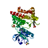
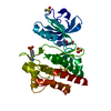
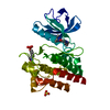
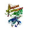
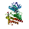
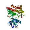
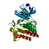
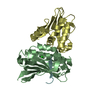
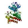
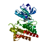
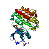
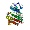
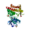
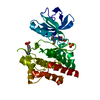
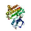
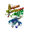
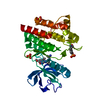
| Title | THE KINASE DOMAIN OF HUMAN LYMPHOCYTE KINASE (LCK), ACTIVATED FORM (AUTO-PHOSPHORYLATED ON TYR394) | ||||||
 Components Components | PROTO-ONCOGENE TYROSINE-PROTEIN KINASE | ||||||
 Keywords Keywords | TYROSINE-PROTEIN KINASE / ATP-BINDING /  PHOSPHORYLATION / SIGNAL TRANSDUCTION PHOSPHORYLATION / SIGNAL TRANSDUCTION | ||||||
| Function / homology |  Function and homology informationregulation of lymphocyte activation / positive regulation of leukocyte cell-cell adhesion / Fc-gamma receptor signaling pathway / CD28 co-stimulation / intracellular zinc ion homeostasis / CD4 receptor binding / FLT3 signaling through SRC family kinases / Nef and signal transduction / Nef Mediated CD4 Down-regulation / Interleukin-2 signaling ...regulation of lymphocyte activation / positive regulation of leukocyte cell-cell adhesion / Fc-gamma receptor signaling pathway / CD28 co-stimulation / intracellular zinc ion homeostasis / CD4 receptor binding / FLT3 signaling through SRC family kinases / Nef and signal transduction / Nef Mediated CD4 Down-regulation / Interleukin-2 signaling / CD28 dependent Vav1 pathway / positive regulation of heterotypic cell-cell adhesion / protein serine/threonine phosphatase activity / Regulation of KIT signaling / CTLA4 inhibitory signaling / phospholipase activator activity / leukocyte migration / positive regulation of T cell receptor signaling pathway / pericentriolar material / Translocation of ZAP-70 to Immunological synapse / Phosphorylation of CD3 and TCR zeta chains / PECAM1 interactions / phospholipase binding / CD28 dependent PI3K/Akt signaling / RHOH GTPase cycle / hemopoiesis / Generation of second messenger molecules / immunological synapse / T cell differentiation / PD-1 signaling / CD8 receptor binding / phosphatidylinositol 3-kinase binding / positive regulation of intrinsic apoptotic signaling pathway / peptidyl-tyrosine autophosphorylation / release of sequestered calcium ion into cytosol / GPVI-mediated activation cascade / T cell costimulation / extrinsic component of cytoplasmic side of plasma membrane / SH2 domain binding / phosphotyrosine residue binding / T cell receptor binding / Signaling by phosphorylated juxtamembrane, extracellular and kinase domain KIT mutants / B cell receptor signaling pathway / non-specific protein-tyrosine kinase / non-membrane spanning protein tyrosine kinase activity / Signaling by SCF-KIT / platelet activation / peptidyl-tyrosine phosphorylation / cell surface receptor protein tyrosine kinase signaling pathway / activation of cysteine-type endopeptidase activity involved in apoptotic process / Constitutive Signaling by Aberrant PI3K in Cancer / positive regulation of T cell activation / Downstream TCR signaling / PIP3 activates AKT signaling / DAP12 signaling / ATPase binding / T cell receptor signaling pathway / PI5P, PP2A and IER3 Regulate PI3K/AKT Signaling / protein phosphatase binding / protein tyrosine kinase activity / intracellular signal transduction / response to xenobiotic stimulus / membrane raft / signaling receptor binding / protein phosphorylation / innate immune response / protein kinase binding / extracellular exosome / ATP binding / identical protein binding / plasma membrane / cytosol Function and homology informationregulation of lymphocyte activation / positive regulation of leukocyte cell-cell adhesion / Fc-gamma receptor signaling pathway / CD28 co-stimulation / intracellular zinc ion homeostasis / CD4 receptor binding / FLT3 signaling through SRC family kinases / Nef and signal transduction / Nef Mediated CD4 Down-regulation / Interleukin-2 signaling ...regulation of lymphocyte activation / positive regulation of leukocyte cell-cell adhesion / Fc-gamma receptor signaling pathway / CD28 co-stimulation / intracellular zinc ion homeostasis / CD4 receptor binding / FLT3 signaling through SRC family kinases / Nef and signal transduction / Nef Mediated CD4 Down-regulation / Interleukin-2 signaling / CD28 dependent Vav1 pathway / positive regulation of heterotypic cell-cell adhesion / protein serine/threonine phosphatase activity / Regulation of KIT signaling / CTLA4 inhibitory signaling / phospholipase activator activity / leukocyte migration / positive regulation of T cell receptor signaling pathway / pericentriolar material / Translocation of ZAP-70 to Immunological synapse / Phosphorylation of CD3 and TCR zeta chains / PECAM1 interactions / phospholipase binding / CD28 dependent PI3K/Akt signaling / RHOH GTPase cycle / hemopoiesis / Generation of second messenger molecules / immunological synapse / T cell differentiation / PD-1 signaling / CD8 receptor binding / phosphatidylinositol 3-kinase binding / positive regulation of intrinsic apoptotic signaling pathway / peptidyl-tyrosine autophosphorylation / release of sequestered calcium ion into cytosol / GPVI-mediated activation cascade / T cell costimulation / extrinsic component of cytoplasmic side of plasma membrane / SH2 domain binding / phosphotyrosine residue binding / T cell receptor binding / Signaling by phosphorylated juxtamembrane, extracellular and kinase domain KIT mutants / B cell receptor signaling pathway / non-specific protein-tyrosine kinase / non-membrane spanning protein tyrosine kinase activity / Signaling by SCF-KIT / platelet activation / peptidyl-tyrosine phosphorylation / cell surface receptor protein tyrosine kinase signaling pathway / activation of cysteine-type endopeptidase activity involved in apoptotic process / Constitutive Signaling by Aberrant PI3K in Cancer / positive regulation of T cell activation / Downstream TCR signaling / PIP3 activates AKT signaling / DAP12 signaling / ATPase binding / T cell receptor signaling pathway / PI5P, PP2A and IER3 Regulate PI3K/AKT Signaling / protein phosphatase binding / protein tyrosine kinase activity / intracellular signal transduction / response to xenobiotic stimulus / membrane raft / signaling receptor binding / protein phosphorylation / innate immune response / protein kinase binding / extracellular exosome / ATP binding / identical protein binding / plasma membrane / cytosolSimilarity search - Function | ||||||
| Biological species |  Homo sapiens (human) Homo sapiens (human) | ||||||
| Method | X-RAY DIFFRACTION / SYNCHROTRON / MIR, MOLECULAR REPLACEMENT, MAD / Resolution: 1.7 Å | ||||||
 Authors Authors | Yamaguchi, H. / Hendrickson, W.A. | ||||||
 Citation Citation | Journal: Nature / Year: 1996 Title: Structural basis for activation of human lymphocyte kinase Lck upon tyrosine phosphorylation. Authors: Yamaguchi, H. / Hendrickson, W.A. | ||||||
| History |
|
- Structure visualization
| Structure viewer | Molecule: MolmilJmol/JSmol |
|---|
- Downloads & links
-Download
| PDBx/mmCIF format | 3lck.cif.gz | 75.2 KB | Display | PDBx/mmCIF format |
|---|---|---|---|---|
| PDB format | pdb3lck.ent.gz | 58.4 KB | Display | PDB format |
| PDBx/mmJSON format | 3lck.json.gz | Tree view | PDBx/mmJSON format | |
| Others |  Other downloads Other downloads |
-Validation report
| Arichive directory | https://data.pdbj.org/pub/pdb/validation_reports/lc/3lckftp://data.pdbj.org/pub/pdb/validation_reports/lc/3lck | HTTPS FTP |
|---|
-Related structure data
| Related structure data |  1irkS S: Starting model for refinement |
|---|---|
| Similar structure data |


-Links
 PDBj
PDBj


- Assembly
| Deposited unit | 
| ||||||||
|---|---|---|---|---|---|---|---|---|---|
| 1 |
| ||||||||
| Unit cell |
|
-Components
| #1: Protein | Mass: 31425.865 Da / Num. of mol.: 1 / Fragment: PROTEIN TYROSINE KINASE DOMAIN Source method: isolated from a genetically manipulated source Details: PHOSPHORYLATION ON TYR 394 / Source: (gene. exp.) Homo sapiens (human) / Cell line: SF9 / Gene: LCK / Plasmid: PFASTBAC-1-BMON14272 / Cell line (production host): SF9 / Cellular location (production host): CYTOPLASM / Gene (production host): POLYHEDRIN / Production host:   Spodoptera frugiperda (fall armyworm) / References: UniProt: P06239, EC: 2.7.1.112 Spodoptera frugiperda (fall armyworm) / References: UniProt: P06239, EC: 2.7.1.112 |
|---|---|
| #2: Chemical | ChemComp-SO4 / Sulfate  Mass: 96.063 Da / Num. of mol.: 1 / Source method: obtained synthetically / Formula: SO4 Mass: 96.063 Da / Num. of mol.: 1 / Source method: obtained synthetically / Formula: SO4 |
| #3: Water | ChemComp-HOH / Water Mass: 18.015 Da / Num. of mol.: 340 / Source method: isolated from a natural source / Formula: H2O Mass: 18.015 Da / Num. of mol.: 340 / Source method: isolated from a natural source / Formula: H2O |
-Experimental details
-Experiment
| Experiment | Method: X-RAY DIFFRACTION / Number of used crystals: 1 |
|---|
- Sample preparation
| Crystal | Density Matthews: 2.14 Å3/Da / Density % sol: 42.5 % | ||||||||||||||||||||||||||||||||||||||||
|---|---|---|---|---|---|---|---|---|---|---|---|---|---|---|---|---|---|---|---|---|---|---|---|---|---|---|---|---|---|---|---|---|---|---|---|---|---|---|---|---|---|
| Crystal grow | Method: vapor diffusion, hanging drop / pH: 6.5 Details: PROTEIN WAS CRYSTALLIZED IN HANGING DROPS WITH 1.6 M AMMONIUM SULFATE AND 0.1 M BISTRIS-HCL (PH 6.5 @ RT) AS A WELL SOLUTION., vapor diffusion - hanging drop Temp details: room temp | ||||||||||||||||||||||||||||||||||||||||
| Crystal grow | *PLUS Method: vapor diffusion, hanging drop / Details: used to seeding | ||||||||||||||||||||||||||||||||||||||||
| Components of the solutions | *PLUS
|
-Data collection
| Diffraction | Mean temperature: 110 K |
|---|---|
| Diffraction source | Source: SYNCHROTRON / Site: NSLS  / Beamline: X4A / Wavelength: 0.9795 / Beamline: X4A / Wavelength: 0.9795 |
| Detector | Type: FUJI / Detector: IMAGE PLATE / Date: Jul 21, 1996 / Details: SAGITTAL FOCUSING MIRROR |
| Radiation | Monochromator: SI(111) / Monochromatic (M) / Laue (L): M / Scattering type: x-ray |
| Radiation wavelength | Wavelength: 0.9795 Å / Relative weight: 1 |
| Reflection | Resolution: 1.7→20 Å / Num. obs: 31599 / % possible obs: 99 % / Redundancy: 8.1 % / Biso Wilson estimate: 13 Å2 / Rsym value: 0.028 / Net I/σ(I): 37 |
| Reflection shell | Resolution: 1.7→1.76 Å / Redundancy: 3.8 % / Mean I/σ(I) obs: 21.8 / Rsym value: 0.047 / % possible all: 96.1 |
| Reflection | *PLUS Rmerge(I) obs: 0.028 |
| Reflection shell | *PLUS Highest resolution: 1.7 Å / % possible obs: 96.1 % / Rmerge(I) obs: 0.047 |
- Processing
| Software |
| ||||||||||||||||||||||||||||||||||||||||||||||||||||||||||||
|---|---|---|---|---|---|---|---|---|---|---|---|---|---|---|---|---|---|---|---|---|---|---|---|---|---|---|---|---|---|---|---|---|---|---|---|---|---|---|---|---|---|---|---|---|---|---|---|---|---|---|---|---|---|---|---|---|---|---|---|---|---|
| Refinement | Method to determine structure: MIR, MOLECULAR REPLACEMENT, MAD Starting model: PDB ENTRY 1IRK Resolution: 1.7→10 Å / Data cutoff high absF: 100000 / Data cutoff low absF: 0.1 / Isotropic thermal model: RESTRAINED Cross valid method: R FREE THROUGHOUT EXCEPT FOR THE LAST ROUND σ(F): 2
| ||||||||||||||||||||||||||||||||||||||||||||||||||||||||||||
| Displacement parameters | Biso mean: 10.37 Å2 | ||||||||||||||||||||||||||||||||||||||||||||||||||||||||||||
| Refinement step | Cycle: LAST / Resolution: 1.7→10 Å
| ||||||||||||||||||||||||||||||||||||||||||||||||||||||||||||
| Refine LS restraints |
| ||||||||||||||||||||||||||||||||||||||||||||||||||||||||||||
| LS refinement shell | Resolution: 1.7→1.78 Å / Total num. of bins used: 8
| ||||||||||||||||||||||||||||||||||||||||||||||||||||||||||||
| Xplor file |
| ||||||||||||||||||||||||||||||||||||||||||||||||||||||||||||
| Software | *PLUS Name: X-PLOR / Version: 3.1 / Classification: refinement | ||||||||||||||||||||||||||||||||||||||||||||||||||||||||||||
| Refine LS restraints | *PLUS
|
