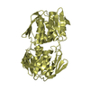
 Movie
Movie Controller


+ Open data

- Basic information
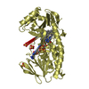
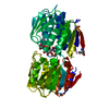
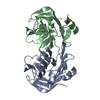
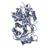
| Entry | Database: PDB / ID: 3kqj | ||||||
|---|---|---|---|---|---|---|---|
| Title | MurA binary complex with UDP-N-acetylglucosamine | ||||||
 Components Components | UDP-N-acetylglucosamine 1-carboxyvinyltransferase | ||||||
 Keywords Keywords | TRANSFERASE / closed enzyme state / inside-out alpha/beta barrel / Cell cycle / Cell division / Cell shape / Cell wall biogenesis/degradation / Peptidoglycan synthesis | ||||||
| Function / homology |  Function and homology informationUDP-N-acetylglucosamine 1-carboxyvinyltransferase / UDP-N-acetylglucosamine 1-carboxyvinyltransferase activity / UDP-N-acetylgalactosamine biosynthetic process / peptidoglycan biosynthetic process / cell wall organization / regulation of cell shape / cell cycle / cell division / cytosol Function and homology informationUDP-N-acetylglucosamine 1-carboxyvinyltransferase / UDP-N-acetylglucosamine 1-carboxyvinyltransferase activity / UDP-N-acetylgalactosamine biosynthetic process / peptidoglycan biosynthetic process / cell wall organization / regulation of cell shape / cell cycle / cell division / cytosolSimilarity search - Function | ||||||
| Biological species |  Escherichia coli (E. coli) Escherichia coli (E. coli) | ||||||
| Method | X-RAY DIFFRACTION / MOLECULAR REPLACEMENT / Resolution: 1.7 Å | ||||||
 Authors Authors | Schonbrunn, E. | ||||||
 Citation Citation | Journal: To be Published Title: The Natural Product Antibiotic Terreic Acid is a Mechanism-Based Inhibitor of the Bacterial Enzyme MurA in vitro but not in vivo. Authors: Han, H. / Yang, Y. / Olesen, S.H. / Becker, A. / Betzi, S. / Schonbrunn, E. | ||||||
| History |
|
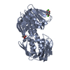
- Structure visualization
| Structure viewer | Molecule: MolmilJmol/JSmol |
|---|
- Downloads & links
-Download
| PDBx/mmCIF format | 3kqj.cif.gz | 105.8 KB | Display | PDBx/mmCIF format |
|---|---|---|---|---|
| PDB format | pdb3kqj.ent.gz | 78.7 KB | Display | PDB format |
| PDBx/mmJSON format | 3kqj.json.gz | Tree view | PDBx/mmJSON format | |
| Others |  Other downloads Other downloads |
-Validation report
| Arichive directory | https://data.pdbj.org/pub/pdb/validation_reports/kq/3kqjftp://data.pdbj.org/pub/pdb/validation_reports/kq/3kqj | HTTPS FTP |
|---|
-Related structure data
| Related structure data | 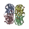 1rywS S: Starting model for refinement |
|---|---|
| Similar structure data |


-Links
 PDBj
PDBj
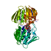
- Assembly
| Deposited unit | 
| ||||||||
|---|---|---|---|---|---|---|---|---|---|
| 1 |
| ||||||||
| Unit cell |
|
-Components
| #1: Protein | / Enoylpyruvate transferase / UDP-N-acetylglucosamine enolpyruvyl transferase / EPT Mass: 44872.523 Da / Num. of mol.: 1 Source method: isolated from a genetically manipulated source Source: (gene. exp.) Escherichia coli (E. coli) / Strain: K12 / Gene: b3189, JW3156, murA, MurA (MurZ), murZ / Plasmid: pET41a / Production host: Escherichia coli (E. coli) / Strain (production host): BL21References: UniProt: P0A749, UDP-N-acetylglucosamine 1-carboxyvinyltransferase |
|---|---|
| #2: Chemical | ChemComp-GOL / Glycerol  Mass: 92.094 Da / Num. of mol.: 1 / Source method: obtained synthetically / Formula: C3H8O3 Mass: 92.094 Da / Num. of mol.: 1 / Source method: obtained synthetically / Formula: C3H8O3 |
| #3: Chemical | ChemComp-UD1 /   Mass: 607.354 Da / Num. of mol.: 1 / Source method: obtained synthetically / Formula: C17H27N3O17P2 Mass: 607.354 Da / Num. of mol.: 1 / Source method: obtained synthetically / Formula: C17H27N3O17P2 |
| #4: Chemical | ChemComp-PO4 / Phosphate  Mass: 94.971 Da / Num. of mol.: 1 / Source method: obtained synthetically / Formula: PO4 Mass: 94.971 Da / Num. of mol.: 1 / Source method: obtained synthetically / Formula: PO4 |
| #5: Water | ChemComp-HOH / Water Mass: 18.015 Da / Num. of mol.: 470 / Source method: isolated from a natural source / Formula: H2O Mass: 18.015 Da / Num. of mol.: 470 / Source method: isolated from a natural source / Formula: H2O |
| Sequence details | ASP67 FORMS AN ISOPEPTIDI |
-Experimental details
-Experiment
| Experiment | Method: X-RAY DIFFRACTION / Number of used crystals: 1 |
|---|
- Sample preparation
| Crystal | Density Matthews: 2.92 Å3/Da / Density % sol: 57.89 % |
|---|---|
| Crystal grow | Temperature: 292 K / Method: vapor diffusion, hanging drop / pH: 7 Details: 12.5 mM Na-formate, 25 mM Na/K phosphate, 10% PEG 3350, pH 7.0, VAPOR DIFFUSION, HANGING DROP, temperature 292K |
-Data collection
| Diffraction | Mean temperature: 93 K |
|---|---|
| Diffraction source | Source: ROTATING ANODE / Type: RIGAKU MICROMAX-007 HF / Wavelength: 1.542 Å |
| Detector | Type: RIGAKU RAXIS HTC / Detector: IMAGE PLATE / Date: Jul 13, 2009 / Details: mirrors |
| Radiation | Monochromator: mirrors / Protocol: SINGLE WAVELENGTH / Monochromatic (M) / Laue (L): M / Scattering type: x-ray |
| Radiation wavelength | Wavelength: 1.542 Å / Relative weight: 1 |
| Reflection | Resolution: 1.7→20 Å / Num. all: 57712 / Num. obs: 57712 / % possible obs: 98.4 % / Observed criterion σ(F): 0 / Observed criterion σ(I): -3 / Redundancy: 5.1 % / Biso Wilson estimate: 17.9 Å2 / Rmerge(I) obs: 0.062 / Net I/σ(I): 15.2 |
| Reflection shell | Resolution: 1.7→1.8 Å / Redundancy: 4.2 % / Rmerge(I) obs: 0.19 / Mean I/σ(I) obs: 6.2 / Num. unique all: 8390 / % possible all: 92.1 |
- Processing
| Software |
| |||||||||||||||||||||||||
|---|---|---|---|---|---|---|---|---|---|---|---|---|---|---|---|---|---|---|---|---|---|---|---|---|---|---|
| Refinement | Method to determine structure: MOLECULAR REPLACEMENT Starting model: 1RYW Resolution: 1.7→20 Å / Cross valid method: THROUGHOUT / σ(F): 0 / σ(I): -3 / Stereochemistry target values: Engh & Huber
| |||||||||||||||||||||||||
| Displacement parameters | Biso mean: 17.5 Å2 | |||||||||||||||||||||||||
| Refine analyze |
| |||||||||||||||||||||||||
| Refinement step | Cycle: LAST / Resolution: 1.7→20 Å
| |||||||||||||||||||||||||
| Refine LS restraints |
| |||||||||||||||||||||||||
| LS refinement shell | Resolution: 1.7→1.81 Å / Rfactor Rfree error: 0.017
|
