 Movie
Movie Controller


+ Open data

- Basic information
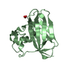
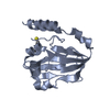
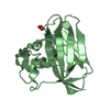
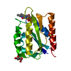
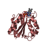
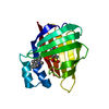
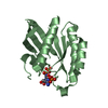
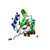
| Entry | Database: PDB / ID: 3klr | ||||||
|---|---|---|---|---|---|---|---|
| Title | Bovine H-protein at 0.88 angstrom resolution | ||||||
 Components Components | Glycine cleavage system H protein | ||||||
 Keywords Keywords | OXIDOREDUCTASE / antiparallel beta sheet / beta sandwich | ||||||
| Function / homology |  Function and homology information Function and homology informationGlycine degradation / Glyoxylate metabolism and glycine degradation / glycine cleavage complex / glycine decarboxylation via glycine cleavage system / mitochondrion / cytoplasmSimilarity search - Function | ||||||
| Biological species |  Bos taurus (cattle) Bos taurus (cattle) | ||||||
| Method | X-RAY DIFFRACTION / SYNCHROTRON / MOLECULAR REPLACEMENT / Resolution: 0.88 Å | ||||||
 Authors Authors | Higashiura, A. / Kurakane, T. / Matsuda, M. / Suzuki, M. / Inaka, K. / Sato, M. / Tanaka, H. / Fujiwara, K. / Nakagawa, A. | ||||||
 Citation Citation | Journal: Acta Crystallogr.,Sect.D / Year: 2010 Title: High-resolution X-ray crystal structure of bovine H-protein at 0.88 A resolution Authors: Higashiura, A. / Kurakane, T. / Matsuda, M. / Suzuki, M. / Inaka, K. / Sato, M. / Kobayashi, T. / Tanaka, T. / Tanaka, H. / Fujiwara, K. / Nakagawa, A. | ||||||
| History |
|
- Structure visualization
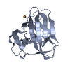
| Structure viewer | Molecule: MolmilJmol/JSmol |
|---|
- Downloads & links
-Download
| PDBx/mmCIF format | 3klr.cif.gz | 83.3 KB | Display | PDBx/mmCIF format |
|---|---|---|---|---|
| PDB format | pdb3klr.ent.gz | 61.7 KB | Display | PDB format |
| PDBx/mmJSON format | 3klr.json.gz | Tree view | PDBx/mmJSON format | |
| Others |  Other downloads Other downloads |
-Validation report
| Arichive directory | https://data.pdbj.org/pub/pdb/validation_reports/kl/3klrftp://data.pdbj.org/pub/pdb/validation_reports/kl/3klr | HTTPS FTP |
|---|
-Related structure data
| Related structure data |  1hpcS S: Starting model for refinement |
|---|---|
| Similar structure data |

-Links
 PDBj
PDBj

- Assembly
| Deposited unit | 
| ||||||||
|---|---|---|---|---|---|---|---|---|---|
| 1 |
| ||||||||
| Unit cell |
| ||||||||
| Components on special symmetry positions |
|
-Components
| #1: Protein | Mass: 13858.369 Da / Num. of mol.: 1 Source method: isolated from a genetically manipulated source Source: (gene. exp.) Bos taurus (cattle) / Plasmid: pET30a / Production host:  Escherichia coli (E. coli) / Strain (production host): BL21(DE3) / References: UniProt: P20821 Escherichia coli (E. coli) / Strain (production host): BL21(DE3) / References: UniProt: P20821 | ||||
|---|---|---|---|---|---|
| #2: Chemical | Sulfate  Mass: 96.063 Da / Num. of mol.: 2 / Source method: obtained synthetically / Formula: SO4 Mass: 96.063 Da / Num. of mol.: 2 / Source method: obtained synthetically / Formula: SO4#3: Chemical | ChemComp-GOL / | Glycerol  Mass: 92.094 Da / Num. of mol.: 1 / Source method: obtained synthetically / Formula: C3H8O3 Mass: 92.094 Da / Num. of mol.: 1 / Source method: obtained synthetically / Formula: C3H8O3#4: Water | ChemComp-HOH / | Water Mass: 18.015 Da / Num. of mol.: 265 / Source method: isolated from a natural source / Formula: H2O Mass: 18.015 Da / Num. of mol.: 265 / Source method: isolated from a natural source / Formula: H2O |
-Experimental details
-Experiment
| Experiment | Method: X-RAY DIFFRACTION / Number of used crystals: 1 |
|---|
- Sample preparation
| Crystal | Density Matthews: 2.72 Å3/Da / Density % sol: 54.72 % / Mosaicity: 0.414 ° |
|---|---|
| Crystal grow | Temperature: 288 K / Method: vapor diffusion, hanging drop / pH: 3 Details: 2.0M ammonium sulfate, 0.1M sodium citrate, pH 3.0, VAPOR DIFFUSION, HANGING DROP, temperature 288K |
-Data collection
| Diffraction | Mean temperature: 100 K |
|---|---|
| Diffraction source | Source: SYNCHROTRON / Site: Photon Factory  / Beamline: BL-5A / Wavelength: 1 Å / Beamline: BL-5A / Wavelength: 1 Å |
| Detector | Type: ADSC QUANTUM 315 / Detector: CCD / Date: Oct 26, 2007 |
| Radiation | Monochromator: Si(111) double crystal monochromator / Protocol: SINGLE WAVELENGTH / Monochromatic (M) / Laue (L): M / Scattering type: x-ray |
| Radiation wavelength | Wavelength: 1 Å / Relative weight: 1 |
| Reflection | Resolution: 0.88→45 Å / Num. obs: 115668 / % possible obs: 98.9 % / Redundancy: 9.6 % / Biso Wilson estimate: 6.95 Å2 / Rmerge(I) obs: 0.055 / Χ2: 1.876 / Net I/σ(I): 19.5 |
| Reflection shell | Resolution: 0.88→0.89 Å / Redundancy: 4.4 % / Rmerge(I) obs: 0.426 / Num. unique all: 3698 / Χ2: 1.089 / % possible all: 95.4 |
- Processing
| Software |
| |||||||||||||||||||||||||||||||||
|---|---|---|---|---|---|---|---|---|---|---|---|---|---|---|---|---|---|---|---|---|---|---|---|---|---|---|---|---|---|---|---|---|---|---|
| Refinement | Method to determine structure: MOLECULAR REPLACEMENT Starting model: PDB ENTRY 1HPC Resolution: 0.88→45 Å / Num. parameters: 12878 / Num. restraintsaints: 16868 / Occupancy max: 1 / Occupancy min: 0 / Cross valid method: THROUGHOUT / σ(I): 4 / Stereochemistry target values: Engh & Huber Details: ANISOTROPIC REFINEMENT REDUCED FREE R (NO CUTOFF) BY ?
| |||||||||||||||||||||||||||||||||
| Solvent computation | Solvent model: MOEWS & KRETSINGER, J.MOL.BIOL.91(1973)201-228 | |||||||||||||||||||||||||||||||||
| Displacement parameters | Biso max: 69.19 Å2 / Biso mean: 14.628 Å2 / Biso min: 5.54 Å2 | |||||||||||||||||||||||||||||||||
| Refinement step | Cycle: LAST / Resolution: 0.88→45 Å
| |||||||||||||||||||||||||||||||||
| Refine LS restraints |
|
