 Movie
Movie Controller


+ Open data

- Basic information
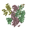
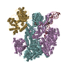
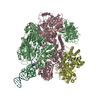
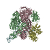
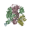
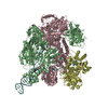
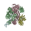
| Entry | Database: PDB / ID: 3k70 | ||||||
|---|---|---|---|---|---|---|---|
| Title | Crystal structure of the complete initiation complex of RecBCD | ||||||
 Components Components |
| ||||||
 Keywords Keywords | HYDROLASE/DNA /  RECOMBINATION / HELICASE / NUCLEASE / HYDROLASE / DNA REPAIR / ATP-binding / DNA damage / Endonuclease / Exonuclease / Nucleotide-binding / HYDROLASE-DNA COMPLEX RECOMBINATION / HELICASE / NUCLEASE / HYDROLASE / DNA REPAIR / ATP-binding / DNA damage / Endonuclease / Exonuclease / Nucleotide-binding / HYDROLASE-DNA COMPLEX | ||||||
| Function / homology |  Function and homology informationexodeoxyribonuclease V / exodeoxyribonuclease V activity / exodeoxyribonuclease V complex / DNA 5'-3' helicase / clearance of foreign intracellular DNA / DNA 3'-5' helicase / DNA translocase activity / single-stranded DNA helicase activity / recombinational repair / 3'-5' DNA helicase activity ...exodeoxyribonuclease V / exodeoxyribonuclease V activity / exodeoxyribonuclease V complex / DNA 5'-3' helicase / clearance of foreign intracellular DNA / DNA 3'-5' helicase / DNA translocase activity / single-stranded DNA helicase activity / recombinational repair / 3'-5' DNA helicase activity / ATP-dependent activity, acting on DNA / DNA helicase activity / DNA endonuclease activity / helicase activity / double-strand break repair via homologous recombination / response to radiation / DNA recombination / DNA damage response / magnesium ion binding / ATP hydrolysis activity / DNA binding / ATP binding / cytosol Function and homology informationexodeoxyribonuclease V / exodeoxyribonuclease V activity / exodeoxyribonuclease V complex / DNA 5'-3' helicase / clearance of foreign intracellular DNA / DNA 3'-5' helicase / DNA translocase activity / single-stranded DNA helicase activity / recombinational repair / 3'-5' DNA helicase activity ...exodeoxyribonuclease V / exodeoxyribonuclease V activity / exodeoxyribonuclease V complex / DNA 5'-3' helicase / clearance of foreign intracellular DNA / DNA 3'-5' helicase / DNA translocase activity / single-stranded DNA helicase activity / recombinational repair / 3'-5' DNA helicase activity / ATP-dependent activity, acting on DNA / DNA helicase activity / DNA endonuclease activity / helicase activity / double-strand break repair via homologous recombination / response to radiation / DNA recombination / DNA damage response / magnesium ion binding / ATP hydrolysis activity / DNA binding / ATP binding / cytosolSimilarity search - Function | ||||||
| Biological species |  Escherichia coli K-12 (bacteria) Escherichia coli K-12 (bacteria) | ||||||
| Method | X-RAY DIFFRACTION / SYNCHROTRON / MOLECULAR REPLACEMENT / Resolution: 3.59 Å | ||||||
 Authors Authors | Saikrishnan, K. / Wigley, D.B. | ||||||
 Citation Citation | Journal: Embo J. / Year: 2008 Title: DNA binding to RecD: role of the 1B domain in SF1B helicase activity. Authors: Saikrishnan, K. / Griffiths, S.P. / Cook, N. / Court, R. / Wigley, D.B. #1: Journal: Nature / Year: 2004Title: Crystal structure of RecBCD enzyme reveals a machine for processing DNA breaks. Authors: Singleton, M.R. / Dillingham, M.S. / Gaudier, M. / Kowalczykowski, S.C. / Wigley, D.B. | ||||||
| History |
|
- Structure visualization
| Structure viewer | Molecule: MolmilJmol/JSmol |
|---|
- Downloads & links
-Download
| PDBx/mmCIF format | 3k70.cif.gz | 1 MB | Display | PDBx/mmCIF format |
|---|---|---|---|---|
| PDB format | pdb3k70.ent.gz | 871.3 KB | Display | PDB format |
| PDBx/mmJSON format | 3k70.json.gz | Tree view | PDBx/mmJSON format | |
| Others |  Other downloads Other downloads |
-Validation report
| Arichive directory | https://data.pdbj.org/pub/pdb/validation_reports/k7/3k70ftp://data.pdbj.org/pub/pdb/validation_reports/k7/3k70 | HTTPS FTP |
|---|
-Related structure data
| Related structure data |  3e1sC  1w36S S: Starting model for refinement C: citing same article ( |
|---|---|
| Similar structure data |


-Links
 PDBj
PDBj


- Assembly
| Deposited unit | 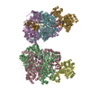
| ||||||||
|---|---|---|---|---|---|---|---|---|---|
| 1 | 
| ||||||||
| 2 | 
| ||||||||
| Unit cell |
| ||||||||
| Details | The quaternary state of the biomolecule is heterotetrameric: the heterotrimeric protein complexes i.e. chain B,C,D and E,F,G, are bound to DNA chain X and Y, respectively. |
-Components
| #1: Protein | Mass: 134110.641 Da / Num. of mol.: 2 Source method: isolated from a genetically manipulated source Source: (gene. exp.) Escherichia coli K-12 (bacteria) / Strain: K12 / Gene: b2820, JW2788, recB, rorA / Production host: Escherichia coli (E. coli) / References: UniProt: P08394, exodeoxyribonuclease V#2: Protein | Mass: 128974.102 Da / Num. of mol.: 2 Source method: isolated from a genetically manipulated source Source: (gene. exp.) Escherichia coli K-12 (bacteria) / Strain: K12 / Gene: b2822, JW2790, recC / Production host: Escherichia coli (E. coli) / References: UniProt: P07648, exodeoxyribonuclease V#3: Protein | Mass: 66990.367 Da / Num. of mol.: 2 Source method: isolated from a genetically manipulated source Source: (gene. exp.) Escherichia coli K-12 (bacteria) / Strain: K12 / Gene: b2819, JW2787, recD / Production host: Escherichia coli (E. coli) / References: UniProt: P04993, exodeoxyribonuclease V#4: DNA chain | Mass: 16628.859 Da / Num. of mol.: 2 / Source method: obtained synthetically / Details: Synthesized DNA #5: Chemical |   Mass: 40.078 Da / Num. of mol.: 2 / Source method: obtained synthetically / Formula: Ca Mass: 40.078 Da / Num. of mol.: 2 / Source method: obtained synthetically / Formula: Ca |
|---|
-Experimental details
-Experiment
| Experiment | Method: X-RAY DIFFRACTION / Number of used crystals: 1 |
|---|
- Sample preparation
| Crystal | Density Matthews: 3.12 Å3/Da / Density % sol: 60.62 % | ||||||||||||||||||||||||||||
|---|---|---|---|---|---|---|---|---|---|---|---|---|---|---|---|---|---|---|---|---|---|---|---|---|---|---|---|---|---|
| Crystal grow | Temperature: 285 K / pH: 7 Details: 100 mM Hepes pH 7.0, 300 mM Calcium acetate, 6-8% PEG 20000, VAPOR DIFFUSION, HANGING DROP, temperature 285.0K | ||||||||||||||||||||||||||||
| Components of the solutions |
|
-Data collection
| Diffraction | Mean temperature: 100 K |
|---|---|
| Diffraction source | Source: SYNCHROTRON / Site: ESRF  / Beamline: ID14-4 / Wavelength: 0.9794 / Beamline: ID14-4 / Wavelength: 0.9794 |
| Detector | Type: ADSC QUANTUM 315r / Detector: CCD / Date: Jul 6, 2006 |
| Radiation | Protocol: SINGLE WAVELENGTH / Monochromatic (M) / Laue (L): M / Scattering type: x-ray |
| Radiation wavelength | Wavelength: 0.9794 Å / Relative weight: 1 |
| Reflection | Resolution: 3.59→50 Å / Num. obs: 97332 / % possible obs: 96.7 % / Observed criterion σ(I): 0 / Redundancy: 3.2 % / Rmerge(I) obs: 0.075 / Rsym value: 0.075 / Net I/σ(I): 6.6 |
| Reflection shell | Resolution: 3.59→3.79 Å / Redundancy: 3.2 % / Rmerge(I) obs: 0.351 / Mean I/σ(I) obs: 2.1 / Rsym value: 0.351 / % possible all: 97.5 |
- Processing
| Software |
| ||||||||||||||||||||||||||||||||||||||||||||||||||||||||||||
|---|---|---|---|---|---|---|---|---|---|---|---|---|---|---|---|---|---|---|---|---|---|---|---|---|---|---|---|---|---|---|---|---|---|---|---|---|---|---|---|---|---|---|---|---|---|---|---|---|---|---|---|---|---|---|---|---|---|---|---|---|---|
| Refinement | Method to determine structure: MOLECULAR REPLACEMENT Starting model: PDB ENTRY 1W36 Resolution: 3.59→30 Å / Cross valid method: THROUGHOUT / σ(F): 0 / Stereochemistry target values: ENGH & HUBER
| ||||||||||||||||||||||||||||||||||||||||||||||||||||||||||||
| Refinement step | Cycle: LAST / Resolution: 3.59→30 Å
| ||||||||||||||||||||||||||||||||||||||||||||||||||||||||||||
| Refine LS restraints |
| ||||||||||||||||||||||||||||||||||||||||||||||||||||||||||||
| LS refinement shell | Resolution: 3.59→3.83 Å
|
