 Movie
Movie Controller

[English] 日本語
 Yorodumi
- PDB-3jsb: Crystal structure of the N-terminal domain of the Lymphocytic Cho... -
+ Open data

- Basic information
| Entry | Database: PDB / ID: 3jsb | ||||||
|---|---|---|---|---|---|---|---|
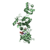
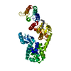
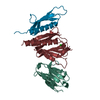
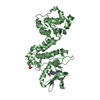
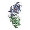
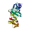
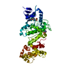
| Title | Crystal structure of the N-terminal domain of the Lymphocytic Choriomeningitis Virus L protein | ||||||
 Components Components | RNA-directed RNA polymerase RNA-dependent RNA polymerase RNA-dependent RNA polymerase | ||||||
 Keywords Keywords | RNA BINDING PROTEIN / VIRAL PROTEIN / VIZIER / Structural Genomics / Marseilles Structural Genomics Program @ AFMB / MSGP / Nucleotide-binding / Nucleotidyltransferase / RNA replication / RNA-directed RNA polymerase / Transferase / Virion | ||||||
| Function / homology |  Function and homology information Function and homology informationRNA-templated viral transcription / negative stranded viral RNA replication / cap snatching / virion component / host cell cytoplasm / Hydrolases; Acting on ester bonds / hydrolase activity / RNA-directed RNA polymerase / RNA-dependent RNA polymerase activity / nucleotide binding / metal ion bindingSimilarity search - Function | ||||||
| Biological species |  Lymphocytic choriomeningitis virus Lymphocytic choriomeningitis virus | ||||||
| Method | X-RAY DIFFRACTION / SYNCHROTRON / MOLECULAR REPLACEMENT / Resolution: 2.13 Å | ||||||
 Authors Authors | Morin, B. / Jamal, S. / Ferron, F.P. / Coutard, B. / Bricogne, G. / Canard, B. / Vonrhein, C. / Marseilles Structural Genomics Program @ AFMB (MSGP) | ||||||
 Citation Citation | Journal: Plos Pathog. / Year: 2010 Title: The N-terminal domain of the arenavirus L protein is an RNA endonuclease essential in mRNA transcription Authors: Morin, B. / Coutard, B. / Lelke, M. / Ferron, F.P. / Kerber, R. / Jamal, S. / Frangeul, A. / Baronti, C. / Charrel, R. / de Lamballerie, X. / Vonrhein, C. / Lescar, J. / Bricogne, G. / Gunther, S. / Canard, B. | ||||||
| History |
|
- Structure visualization
| Structure viewer | Molecule: MolmilJmol/JSmol |
|---|
- Downloads & links
-Download
| PDBx/mmCIF format | 3jsb.cif.gz | 170 KB | Display | PDBx/mmCIF format |
|---|---|---|---|---|
| PDB format | pdb3jsb.ent.gz | 136.8 KB | Display | PDB format |
| PDBx/mmJSON format | 3jsb.json.gz | Tree view | PDBx/mmJSON format | |
| Others |  Other downloads Other downloads |
-Validation report
| Arichive directory | https://data.pdbj.org/pub/pdb/validation_reports/js/3jsbftp://data.pdbj.org/pub/pdb/validation_reports/js/3jsb | HTTPS FTP |
|---|
-Related structure data
| Similar structure data |
|---|


-Links
 PDBj
PDBj- Assembly
| Deposited unit | 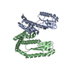
| ||||||||
|---|---|---|---|---|---|---|---|---|---|
| 1 | 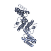
| ||||||||
| 2 | 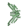
| ||||||||
| Unit cell |
|
-Components
| #1: Protein | RNA-dependent RNA polymerase / Protein L Mass: 23722.172 Da / Num. of mol.: 2 / Fragment: UNP residues 2-197 / Mutation: S107T, N173D Source method: isolated from a genetically manipulated source Source: (gene. exp.) Lymphocytic choriomeningitis virus / Strain: Armstrong / Gene: L / Plasmid: pDest14 / Production host:  Escherichia coli (E. coli) / Strain (production host): T7 express Iq (biolabs-C3016) / References: UniProt: P14240, RNA-directed RNA polymerase Escherichia coli (E. coli) / Strain (production host): T7 express Iq (biolabs-C3016) / References: UniProt: P14240, RNA-directed RNA polymerase#2: Water | ChemComp-HOH / | Water Mass: 18.015 Da / Num. of mol.: 252 / Source method: isolated from a natural source / Formula: H2O Mass: 18.015 Da / Num. of mol.: 252 / Source method: isolated from a natural source / Formula: H2O |
|---|
-Experimental details
-Experiment
| Experiment | Method: X-RAY DIFFRACTION / Number of used crystals: 1 |
|---|
- Sample preparation
| Crystal | Density Matthews: 3.21 Å3/Da / Density % sol: 61.71 % |
|---|---|
| Crystal grow | Temperature: 293 K / Method: vapor diffusion, sitting drop / pH: 5 Details: 0.2M-0.8M LiSo4, 50mM Citrate, 5-10% Isopropanol, VAPOR DIFFUSION, SITTING DROP, temperature 293K |
-Data collection
| Diffraction | Mean temperature: 200 K |
|---|---|
| Diffraction source | Source: SYNCHROTRON / Site: ESRF  / Beamline: ID14-4 / Wavelength: 0.9835 Å / Beamline: ID14-4 / Wavelength: 0.9835 Å |
| Detector | Type: ADSC QUANTUM 315r / Detector: CCD / Date: Jul 29, 2008 |
| Radiation | Protocol: SINGLE WAVELENGTH / Monochromatic (M) / Laue (L): M / Scattering type: x-ray |
| Radiation wavelength | Wavelength: 0.9835 Å / Relative weight: 1 |
| Reflection | Resolution: 2.13→107.25 Å / Num. obs: 33999 / % possible obs: 97.9 % / Observed criterion σ(F): 0 / Observed criterion σ(I): 0 / Redundancy: 6.8 % / Biso Wilson estimate: 48.62 Å2 / Rmerge(I) obs: 0.039 / Rsym value: 0.039 / Net I/σ(I): 27.1 / Num. measured all: 215459 |
| Reflection shell | Resolution: 2.13→2.19 Å / Redundancy: 2.9 % / Mean I/σ(I) obs: 1.9 / % possible all: 97.8 |
- Processing
| Software |
| ||||||||||||||||||||||||||||||||||||||||||||||||||||||||||||||||||||||||||||||||||||||||||||||||||||||||||||||||||
|---|---|---|---|---|---|---|---|---|---|---|---|---|---|---|---|---|---|---|---|---|---|---|---|---|---|---|---|---|---|---|---|---|---|---|---|---|---|---|---|---|---|---|---|---|---|---|---|---|---|---|---|---|---|---|---|---|---|---|---|---|---|---|---|---|---|---|---|---|---|---|---|---|---|---|---|---|---|---|---|---|---|---|---|---|---|---|---|---|---|---|---|---|---|---|---|---|---|---|---|---|---|---|---|---|---|---|---|---|---|---|---|---|---|---|---|
| Refinement | Method to determine structure: MOLECULAR REPLACEMENT / Resolution: 2.13→42.59 Å / Cor.coef. Fo:Fc: 0.9405 / Cor.coef. Fo:Fc free: 0.9353 / Occupancy max: 1 / Occupancy min: 0.11 / SU B: 5.865 / SU ML: 0.151 / SU R Cruickshank DPI: 0.168 / Cross valid method: THROUGHOUT / σ(F): 0 / ESU R: 0.241 / ESU R Free: 0.21 / SU R Blow DPI: 0.176 / SU Rfree Blow DPI: 0.15 / SU Rfree Cruickshank DPI: 0.147 / Stereochemistry target values: MAXIMUM LIKELIHOOD / Details: HYDROGENS HAVE BEEN ADDED IN THE RIDING POSITIONS
| ||||||||||||||||||||||||||||||||||||||||||||||||||||||||||||||||||||||||||||||||||||||||||||||||||||||||||||||||||
| Solvent computation | Ion probe radii: 0.8 Å / Shrinkage radii: 0.8 Å / VDW probe radii: 1.4 Å / Solvent model: MASK | ||||||||||||||||||||||||||||||||||||||||||||||||||||||||||||||||||||||||||||||||||||||||||||||||||||||||||||||||||
| Displacement parameters | Biso max: 144.99 Å2 / Biso mean: 59.853 Å2 / Biso min: 27.28 Å2
| ||||||||||||||||||||||||||||||||||||||||||||||||||||||||||||||||||||||||||||||||||||||||||||||||||||||||||||||||||
| Refine analyze | Luzzati coordinate error obs: 0.297 Å | ||||||||||||||||||||||||||||||||||||||||||||||||||||||||||||||||||||||||||||||||||||||||||||||||||||||||||||||||||
| Refinement step | Cycle: LAST / Resolution: 2.13→42.59 Å
| ||||||||||||||||||||||||||||||||||||||||||||||||||||||||||||||||||||||||||||||||||||||||||||||||||||||||||||||||||
| Refine LS restraints |
| ||||||||||||||||||||||||||||||||||||||||||||||||||||||||||||||||||||||||||||||||||||||||||||||||||||||||||||||||||
| LS refinement shell | Resolution: 2.13→2.19 Å / Total num. of bins used: 17
| ||||||||||||||||||||||||||||||||||||||||||||||||||||||||||||||||||||||||||||||||||||||||||||||||||||||||||||||||||
| Refinement TLS params. | Method: refined / Refine-ID: X-RAY DIFFRACTION
| ||||||||||||||||||||||||||||||||||||||||||||||||||||||||||||||||||||||||||||||||||||||||||||||||||||||||||||||||||
| Refinement TLS group |
|
