 Movie
Movie Controller


+ Open data

- Basic information
| Entry | Database: PDB / ID: 3jam | ||||||
|---|---|---|---|---|---|---|---|
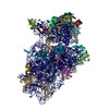
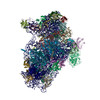
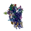
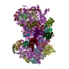
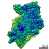
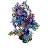
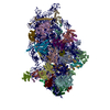
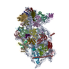
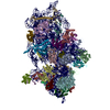
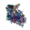
| Title | CryoEM structure of 40S-eIF1A-eIF1 complex from yeast | ||||||
 Components Components |
| ||||||
 Keywords Keywords |  TRANSLATION / Eukaryotic translation initiation / 48S / small ribosome subunit / 40S / 43S TRANSLATION / Eukaryotic translation initiation / 48S / small ribosome subunit / 40S / 43S | ||||||
| Function / homology |  Function and homology information Function and homology informationformation of translation initiation ternary complex / translation reinitiation / multi-eIF complex / eukaryotic 43S preinitiation complex / formation of cytoplasmic translation initiation complex / formation of translation preinitiation complex / eukaryotic 48S preinitiation complex / Formation of the ternary complex, and subsequently, the 43S complex / Translation initiation complex formation / Ribosomal scanning and start codon recognition ...formation of translation initiation ternary complex / translation reinitiation / multi-eIF complex / eukaryotic 43S preinitiation complex / formation of cytoplasmic translation initiation complex / formation of translation preinitiation complex / eukaryotic 48S preinitiation complex / Formation of the ternary complex, and subsequently, the 43S complex / Translation initiation complex formation / Ribosomal scanning and start codon recognition / Formation of a pool of free 40S subunits / ribosomal small subunit binding / L13a-mediated translational silencing of Ceruloplasmin expression / translation regulator activity / regulation of translational fidelity / translation initiation factor binding / translational initiation / translation initiation factor activity / cytosolic ribosome / maintenance of translational fidelity / ribosomal small subunit assembly / rRNA processing / cytoplasmic stress granule / cytosolic small ribosomal subunit / ribosome binding / double-stranded RNA binding / small ribosomal subunit / cytosolic large ribosomal subunit / cytoplasmic translation / rRNA binding / ribosome / structural constituent of ribosome / translation / ribonucleoprotein complex / protein kinase binding / RNA binding / zinc ion binding / metal ion binding / nucleus / cytosol / cytoplasmSimilarity search - Function | ||||||
| Biological species |  Saccharomyces cerevisiae (brewer's yeast)Kluyveromyces lactis (yeast) Saccharomyces cerevisiae (brewer's yeast)Kluyveromyces lactis (yeast) | ||||||
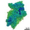
| Method | ELECTRON MICROSCOPY / single particle reconstruction / cryo EM / Resolution: 3.46 Å | ||||||
 Authors Authors | Llacer, J.L. / Hussain, T. / Ramakrishnan, V. | ||||||
 Citation Citation | Journal: Mol Cell / Year: 2015 Title: Conformational Differences between Open and Closed States of the Eukaryotic Translation Initiation Complex. Authors: Jose L Llácer / Tanweer Hussain / Laura Marler / Colin Echeverría Aitken / Anil Thakur / Jon R Lorsch / Alan G Hinnebusch / V Ramakrishnan /   Abstract: Translation initiation in eukaryotes begins with the formation of a pre-initiation complex (PIC) containing the 40S ribosomal subunit, eIF1, eIF1A, eIF3, ternary complex (eIF2-GTP-Met-tRNAi), and ...Translation initiation in eukaryotes begins with the formation of a pre-initiation complex (PIC) containing the 40S ribosomal subunit, eIF1, eIF1A, eIF3, ternary complex (eIF2-GTP-Met-tRNAi), and eIF5. The PIC, in an open conformation, attaches to the 5' end of the mRNA and scans to locate the start codon, whereupon it closes to arrest scanning. We present single particle cryo-electron microscopy (cryo-EM) reconstructions of 48S PICs from yeast in these open and closed states, at 6.0 Å and 4.9 Å, respectively. These reconstructions show eIF2β as well as a configuration of eIF3 that appears to encircle the 40S, occupying part of the subunit interface. Comparison of the complexes reveals a large conformational change in the 40S head from an open mRNA latch conformation to a closed one that constricts the mRNA entry channel and narrows the P site to enclose tRNAi, thus elucidating key events in start codon recognition. | ||||||
| History |
|
- Structure visualization
| Movie |
Movie viewer |
|---|---|
| Structure viewer | Molecule: MolmilJmol/JSmol |
- Downloads & links
-Download
| PDBx/mmCIF format | 3jam.cif.gz | 1.9 MB | Display | PDBx/mmCIF format |
|---|---|---|---|---|
| PDB format | pdb3jam.ent.gz | 1.5 MB | Display | PDB format |
| PDBx/mmJSON format | 3jam.json.gz | Tree view | PDBx/mmJSON format | |
| Others |  Other downloads Other downloads |
-Validation report
| Arichive directory | https://data.pdbj.org/pub/pdb/validation_reports/ja/3jamftp://data.pdbj.org/pub/pdb/validation_reports/ja/3jam | HTTPS FTP |
|---|
-Related structure data
| Related structure data |  3047MC 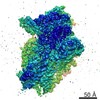 3048C 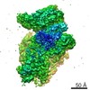 3049C  3050C 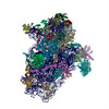 3japC M: map data used to model this data C: citing same article ( |
|---|---|
| Similar structure data |

-Links
 PDBj
PDBj


- Assembly
| Deposited unit | 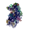
|
|---|---|
| 1 |
|
-Components
+Protein , 35 types, 35 molecules ABCDEFGHIJKLMNOPQRSTUVWXYZabcd...

-RNA chain / Protein/peptide , 2 types, 2 molecules 2h
| #1: RNA chain | 18S ribosomal RNA Mass: 579545.875 Da / Num. of mol.: 1 / Source method: isolated from a natural source / Source: (natural) Kluyveromyces lactis (yeast) / References: GenBank: NC_006040.1 |
|---|---|
| #35: Protein/peptide | Mass: 3354.243 Da / Num. of mol.: 1 / Source method: isolated from a natural source / Source: (natural) Kluyveromyces lactis (yeast) / References: UniProt: P0CX86 |
-Non-polymers , 2 types, 83 molecules 

| #38: Chemical | ChemComp-MG /  Mass: 24.305 Da / Num. of mol.: 80 / Source method: obtained synthetically / Formula: Mg Mass: 24.305 Da / Num. of mol.: 80 / Source method: obtained synthetically / Formula: Mg#39: Chemical |  Mass: 65.409 Da / Num. of mol.: 3 / Source method: obtained synthetically / Formula: Zn Mass: 65.409 Da / Num. of mol.: 3 / Source method: obtained synthetically / Formula: Zn |
|---|
-Experimental details
-Experiment
| Experiment | Method: ELECTRON MICROSCOPY |
|---|---|
| EM experiment | Aggregation state: PARTICLE / 3D reconstruction method: single particle reconstruction |
- Sample preparation
| Component |
| ||||||||||||||||||||
|---|---|---|---|---|---|---|---|---|---|---|---|---|---|---|---|---|---|---|---|---|---|
| Buffer solution | Name: 20 mM MES-KOH, 40 mM potassium acetate, 10 mM ammonium acetate, 8 mM magnesium acetate, 2 mM DTT pH: 6.5 Details: 20 mM MES-KOH, 40 mM potassium acetate, 10 mM ammonium acetate, 8 mM magnesium acetate, 2 mM DTT | ||||||||||||||||||||
| Specimen | Conc.: 0.17 mg/ml / Embedding applied: NO / Shadowing applied: NO / Staining applied: NO / Vitrification applied: YES | ||||||||||||||||||||
| Specimen support | Details: Quantifoil R2/2 400 mesh copper grids with 4-5 nm thin carbon on top | ||||||||||||||||||||
| Vitrification | Instrument: FEI VITROBOT MARK I / Cryogen name: ETHANE / Temp: 120 K / Humidity: 100 % Details: Blot for 2.5 to 3 seconds before plunging into liquid ethane (FEI VITROBOT MARK I). Method: Blot for 2.5 to 3 seconds before plunging |
- Electron microscopy imaging
| Experimental equipment |  Model: Titan Krios / Image courtesy: FEI Company |
|---|---|
| Microscopy | Model: FEI TITAN KRIOS / Date: Apr 28, 2014 Details: Complete dataset was collected over two non-consecutive days. |
| Electron gun | Electron source: FIELD EMISSION GUN / Accelerating voltage: 300 kV / Illumination mode: FLOOD BEAM |
| Electron lens | Mode: BRIGHT FIELDBright-field microscopy / Nominal magnification: 78000 X / Calibrated magnification: 104478 X / Nominal defocus max: 4000 nm / Nominal defocus min: 1500 nm / Cs: 2.7 mm |
| Specimen holder | Specimen holder type: GATAN LIQUID NITROGEN |
| Image recording | Electron dose: 27 e/Å2 / Film or detector model: FEI FALCON II (4k x 4k) |
| Image scans | Num. digital images: 2056 |
| Radiation wavelength | Relative weight: 1 |
- Processing
| Software | Name: REFMAC / Version: 5.8.0124 2015/06/03 / Classification: refinement / Contact author: Garib N. Murshudov / Contact author email: garib[at]mrc-lmb.cam.ac.uk Description: (un)restrained refinement or idealisation of macromolecular structures | |||||||||||||||
|---|---|---|---|---|---|---|---|---|---|---|---|---|---|---|---|---|
| EM software |
| |||||||||||||||
| Symmetry | Point symmetry: C1 (asymmetric) | |||||||||||||||
| 3D reconstruction | Resolution: 3.46 Å / Resolution method: FSC 0.143 CUT-OFF / Num. of particles: 86055 / Nominal pixel size: 1.34 Å / Actual pixel size: 1.34 Å / Details: gold-standard / Refinement type: HALF-MAPS REFINED INDEPENDENTLY / Symmetry type: POINT | |||||||||||||||
| Atomic model building | B value: 89 / Protocol: OTHER / Space: RECIPROCAL / Target criteria: R-factor, FSC / Details: METHOD--Local refinement | |||||||||||||||
| Atomic model building | PDB-ID: 3J80 Accession code: 3J80 / Source name: PDB / Type: experimental model | |||||||||||||||
| Refinement | Details: Hydrogens have been added in their riding positions | |||||||||||||||
| Refinement step | Cycle: LAST
|
