 Movie
Movie Controller

[English] 日本語
 Yorodumi
- PDB-3j3w: Atomic model of the immature 50S subunit from Bacillus subtilis (... -
+ Open data

- Basic information
| Entry | Database: PDB / ID: 3j3w | ||||||
|---|---|---|---|---|---|---|---|
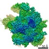
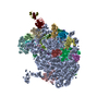
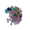
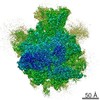
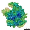
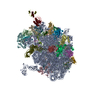
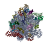
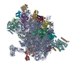
| Title | Atomic model of the immature 50S subunit from Bacillus subtilis (state II-a) | ||||||
 Components Components |
| ||||||
 Keywords Keywords |  RIBOSOME / Ribosome biogenesis / ribosome assembly / RNA folding / YlqF RIBOSOME / Ribosome biogenesis / ribosome assembly / RNA folding / YlqF | ||||||
| Function / homology |  Function and homology information Function and homology informationpositive regulation of rRNA processing / nucleoid / rRNA processing / large ribosomal subunit rRNA binding / large ribosomal subunit / regulation of translation / cytoplasmic translation / cytosolic large ribosomal subunit / transferase activity / tRNA binding ...positive regulation of rRNA processing / nucleoid / rRNA processing / large ribosomal subunit rRNA binding / large ribosomal subunit / regulation of translation / cytoplasmic translation / cytosolic large ribosomal subunit / transferase activity / tRNA binding / negative regulation of translation / rRNA binding / ribosome / structural constituent of ribosome / ribonucleoprotein complex / translation / response to antibiotic / mRNA binding / DNA binding / RNA binding / cytoplasmSimilarity search - Function | ||||||
| Biological species |  Bacillus subtilis (bacteria) Bacillus subtilis (bacteria) | ||||||
| Method | ELECTRON MICROSCOPY / single particle reconstruction / cryo EM / Resolution: 10.7 Å | ||||||
 Authors Authors | Li, N. / Guo, Q. / Zhang, Y. / Yuan, Y. / Ma, C. / Lei, J. / Gao, N. | ||||||
 Citation Citation | Journal: Nucleic Acids Res / Year: 2013 Title: Cryo-EM structures of the late-stage assembly intermediates of the bacterial 50S ribosomal subunit. Authors: Ningning Li / Yuling Chen / Qiang Guo / Yixiao Zhang / Yi Yuan / Chengying Ma / Haiteng Deng / Jianlin Lei / Ning Gao /  Abstract: Ribosome assembly is a process fundamental for all cellular activities. The efficiency and accuracy of the subunit assembly are tightly regulated and closely monitored. In the present work, we ...Ribosome assembly is a process fundamental for all cellular activities. The efficiency and accuracy of the subunit assembly are tightly regulated and closely monitored. In the present work, we characterized, both compositionally and structurally, a set of in vivo 50S subunit precursors (45S), isolated from a mutant bacterial strain. Our qualitative mass spectrometry data indicate that L28, L16, L33, L36 and L35 are dramatically underrepresented in the 45S particles. This protein spectrum shows interesting similarity to many qualitatively analyzed 50S precursors from different genetic background, indicating the presence of global rate-limiting steps in the late-stage assembly of 50S subunit. Our structural data reveal two major intermediate states for the 45S particles. Consistently, both states severally lack those proteins, but they also differ in the stability of the functional centers of the 50S subunit, demonstrating that they are translationally inactive. Detailed analysis indicates that the orientation of H38 accounts for the global conformational differences in these intermediate structures, and suggests that the reorientation of H38 to its native position is rate-limiting during the late-stage assembly. Especially, H38 plays an essential role in stabilizing the central protuberance, through the interaction with the 5S rRNA, and the correctly orientated H38 is likely a prerequisite for further maturation of the 50S subunit. | ||||||
| History |
|
- Structure visualization
| Movie |
Movie viewer |
|---|---|
| Structure viewer | Molecule: MolmilJmol/JSmol |
- Downloads & links
-Download
| PDBx/mmCIF format | 3j3w.cif.gz | 1.7 MB | Display | PDBx/mmCIF format |
|---|---|---|---|---|
| PDB format | pdb3j3w.ent.gz | 1.2 MB | Display | PDB format |
| PDBx/mmJSON format | 3j3w.json.gz | Tree view | PDBx/mmJSON format | |
| Others |  Other downloads Other downloads |
-Validation report
| Arichive directory | https://data.pdbj.org/pub/pdb/validation_reports/j3/3j3wftp://data.pdbj.org/pub/pdb/validation_reports/j3/3j3w | HTTPS FTP |
|---|
-Related structure data
| Related structure data | 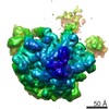 5643MC 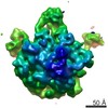 5642C 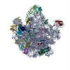 3j3vC M: map data used to model this data C: citing same article ( |
|---|---|
| Similar structure data |

-Links
 PDBj
PDBj


- Assembly
| Deposited unit | 
|
|---|---|
| 1 |
|
-Components
-RNA chain , 1 types, 1 molecules A
| #1: RNA chain | Mass: 949324.125 Da / Num. of mol.: 1 / Source method: isolated from a natural source / Source: (natural) Bacillus subtilis (bacteria) / Strain: 168 / References: GenBank: AL009126.3 |
|---|
-50S ribosomal protein ... , 19 types, 19 molecules 0CNGJKLPQDRSTUX256E
| #2: Protein | Mass: 6745.073 Da / Num. of mol.: 1 / Source method: isolated from a natural source / Source: (natural) Bacillus subtilis (bacteria) / Strain: 168 / References: UniProt: O34687 |
|---|---|
| #3: Protein | / BL2 Mass: 30335.125 Da / Num. of mol.: 1 / Source method: isolated from a natural source / Source: (natural) Bacillus subtilis (bacteria) / Strain: 168 / References: UniProt: P42919 |
| #4: Protein | / BL15 / BL21 Mass: 13774.806 Da / Num. of mol.: 1 / Source method: isolated from a natural source / Source: (natural) Bacillus subtilis (bacteria) / Strain: 168 / References: UniProt: P20277 |
| #5: Protein | / BL10 Mass: 19543.389 Da / Num. of mol.: 1 / Source method: isolated from a natural source / Source: (natural) Bacillus subtilis (bacteria) / Strain: 168 / References: UniProt: P46898 |
| #6: Protein | Mass: 16407.104 Da / Num. of mol.: 1 / Source method: isolated from a natural source / Source: (natural) Bacillus subtilis (bacteria) / Strain: 168 / References: UniProt: P70974 |
| #7: Protein | Mass: 13175.288 Da / Num. of mol.: 1 / Source method: isolated from a natural source / Source: (natural) Bacillus subtilis (bacteria) / Strain: 168 / References: UniProt: P12875 |
| #8: Protein | Mass: 15410.694 Da / Num. of mol.: 1 / Source method: isolated from a natural source / Source: (natural) Bacillus subtilis (bacteria) / Strain: 168 / References: UniProt: P19946 |
| #9: Protein | Mass: 13416.853 Da / Num. of mol.: 1 / Source method: isolated from a natural source / Source: (natural) Bacillus subtilis (bacteria) / Strain: 168 / References: UniProt: O31742 |
| #10: Protein | Mass: 13669.189 Da / Num. of mol.: 1 / Source method: isolated from a natural source / Source: (natural) Bacillus subtilis (bacteria) / Strain: 168 / References: UniProt: P55873 |
| #11: Protein | / BL3 Mass: 22723.348 Da / Num. of mol.: 1 / Source method: isolated from a natural source / Source: (natural) Bacillus subtilis (bacteria) / Strain: 168 / References: UniProt: P42920 |
| #12: Protein | / BL20 Mass: 11296.081 Da / Num. of mol.: 1 / Source method: isolated from a natural source / Source: (natural) Bacillus subtilis (bacteria) / Strain: 168 / References: UniProt: P26908 |
| #13: Protein | Mass: 12481.608 Da / Num. of mol.: 1 / Source method: isolated from a natural source / Source: (natural) Bacillus subtilis (bacteria) / Strain: 168 / References: UniProt: P42060 |
| #14: Protein | Mass: 10978.813 Da / Num. of mol.: 1 / Source method: isolated from a natural source / Source: (natural) Bacillus subtilis (bacteria) / Strain: 168 / References: UniProt: P42924 |
| #15: Protein | / 12 kDa DNA-binding protein / BL23 / HPB12 Mass: 11166.120 Da / Num. of mol.: 1 / Source method: isolated from a natural source / Source: (natural) Bacillus subtilis (bacteria) / Strain: 168 / References: UniProt: P0CI78 |
| #16: Protein | Mass: 7728.029 Da / Num. of mol.: 1 / Source method: isolated from a natural source / Source: (natural) Bacillus subtilis (bacteria) / Strain: 168 / References: UniProt: P12873 |
| #17: Protein/peptide | Mass: 5271.332 Da / Num. of mol.: 1 / Source method: isolated from a natural source / Source: (natural) Bacillus subtilis (bacteria) / Strain: 168 / References: UniProt: P05647 |
| #18: Protein | / BL1 Mass: 25026.887 Da / Num. of mol.: 1 / Source method: isolated from a natural source / Source: (natural) Bacillus subtilis (bacteria) / Strain: 168 / References: UniProt: Q06797 |
| #19: Protein | / BL11 Mass: 14951.442 Da / Num. of mol.: 1 / Source method: isolated from a natural source / Source: (natural) Bacillus subtilis (bacteria) / Strain: 168 / References: UniProt: Q06796 |
| #20: Protein | Mass: 22424.951 Da / Num. of mol.: 1 / Source method: isolated from a natural source / Source: (natural) Bacillus subtilis (bacteria) / Strain: 168 / References: UniProt: P42921 |
-Experimental details
-Experiment
| Experiment | Method: ELECTRON MICROSCOPY |
|---|---|
| EM experiment | Aggregation state: PARTICLE / 3D reconstruction method: single particle reconstruction |
- Sample preparation
| Component | Name: Immature 50S subunit from YlqF-deficient Bacillus subtilis strain Type: RIBOSOME |
|---|---|
| Buffer solution | Name: 100mM NH4Cl, 20mM Tris-HCl, 10mM MgOAc2, 1mM TCEP / pH: 7.5 / Details: 100mM NH4Cl, 20mM Tris-HCl, 10mM MgOAc2, 1mM TCEP |
| Specimen | Embedding applied: NO / Shadowing applied: NO / Staining applied: NO / Vitrification applied: YES |
| Vitrification | Instrument: FEI VITROBOT MARK IV / Cryogen name: ETHANE / Humidity: 100 % / Method: Blot for 20 seconds before plunging |
- Electron microscopy imaging
| Experimental equipment |  Model: Titan Krios / Image courtesy: FEI Company |
|---|---|
| Microscopy | Model: FEI TITAN KRIOS / Date: Dec 6, 2011 |
| Electron gun | Electron source: FIELD EMISSION GUN / Accelerating voltage: 300 kV / Illumination mode: FLOOD BEAM |
| Electron lens | Mode: BRIGHT FIELDBright-field microscopy / Nominal magnification: 59000 X / Nominal defocus max: 4000 nm / Nominal defocus min: 1000 nm / Cs: 2.7 mm / Camera length: 0 mm |
| Specimen holder | Specimen holder model: FEI TITAN KRIOS AUTOGRID HOLDER / Tilt angle max: 0 ° / Tilt angle min: 0 ° |
| Image recording | Electron dose: 20 e/Å2 / Film or detector model: FEI EAGLE (4k x 4k) |
| Radiation wavelength | Relative weight: 1 |
- Processing
| EM software |
| |||||||||||||||||||||
|---|---|---|---|---|---|---|---|---|---|---|---|---|---|---|---|---|---|---|---|---|---|---|
| CTF correction | Details: Each particle | |||||||||||||||||||||
| Symmetry | Point symmetry: C1 (asymmetric) | |||||||||||||||||||||
| 3D reconstruction | Method: Reference projections / Resolution: 10.7 Å / Resolution method: OTHER / Num. of particles: 27652 Details: Single particle details: This is one of the classified groups with the software RELION (Single particle--Applied symmetry: C1) Symmetry type: POINT | |||||||||||||||||||||
| Atomic model building |
| |||||||||||||||||||||
| Atomic model building |
| |||||||||||||||||||||
| Refinement step | Cycle: LAST
|

