 Movie
Movie Controller

[English] 日本語
 Yorodumi
- PDB-3ilk: The structure of a probable methylase family protein from Haemoph... -
+ Open data

- Basic information
| Entry | Database: PDB / ID: 3ilk | ||||||
|---|---|---|---|---|---|---|---|
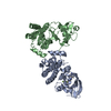
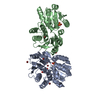
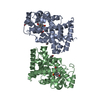
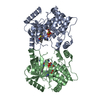
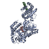
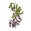
| Title | The structure of a probable methylase family protein from Haemophilus influenzae Rd KW20 | ||||||
 Components Components | Uncharacterized tRNA/rRNA methyltransferase HI0380 | ||||||
 Keywords Keywords |  structural genomics / unknown function / APC63004 / methylase family protein / Haemophilus influenzae Rd KW20 / PSI-2 / protein structure initiative / midwest center for structural genomics / MCSG / Methyltransferase / Transferase structural genomics / unknown function / APC63004 / methylase family protein / Haemophilus influenzae Rd KW20 / PSI-2 / protein structure initiative / midwest center for structural genomics / MCSG / Methyltransferase / Transferase | ||||||
| Function / homology |  Function and homology information: / tRNA nucleoside ribose methylation / tRNA (uracil-2'-O-)-methyltransferase activity / Transferases; Transferring one-carbon groups; Methyltransferases / RNA binding / cytosol Function and homology information: / tRNA nucleoside ribose methylation / tRNA (uracil-2'-O-)-methyltransferase activity / Transferases; Transferring one-carbon groups; Methyltransferases / RNA binding / cytosolSimilarity search - Function | ||||||
| Biological species |  Haemophilus influenzae (bacteria) Haemophilus influenzae (bacteria) | ||||||
| Method | X-RAY DIFFRACTION / SYNCHROTRON / SAD / Resolution: 2.01 Å | ||||||
 Authors Authors | Tan, K. / Li, H. / Buck, K. / Joachimiak, A. / Midwest Center for Structural Genomics (MCSG) | ||||||
 Citation Citation | Journal: To be Published Title: The structure of a probable methylase family protein from Haemophilus influenzae Rd KW20 Authors: Tan, K. / Li, H. / Buck, K. / Joachimiak, A. | ||||||
| History |
|
- Structure visualization
| Structure viewer | Molecule: MolmilJmol/JSmol |
|---|
- Downloads & links
-Download
| PDBx/mmCIF format | 3ilk.cif.gz | 104.4 KB | Display | PDBx/mmCIF format |
|---|---|---|---|---|
| PDB format | pdb3ilk.ent.gz | 86.2 KB | Display | PDB format |
| PDBx/mmJSON format | 3ilk.json.gz | Tree view | PDBx/mmJSON format | |
| Others |  Other downloads Other downloads |
-Validation report
| Arichive directory | https://data.pdbj.org/pub/pdb/validation_reports/il/3ilkftp://data.pdbj.org/pub/pdb/validation_reports/il/3ilk | HTTPS FTP |
|---|
-Related structure data
| Similar structure data | |
|---|---|
| Other databases |


-Links
 PDBj
PDBj- Assembly
| Deposited unit | 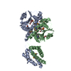
| ||||||||
|---|---|---|---|---|---|---|---|---|---|
| 1 |
| ||||||||
| Unit cell |
| ||||||||
| Details | Authors state that the biological unit is experimentally unknown. The chains A and B likely form a dimer. |
-Components
| #1: Protein | Mass: 27875.916 Da / Num. of mol.: 2 Source method: isolated from a genetically manipulated source Source: (gene. exp.) Haemophilus influenzae (bacteria) / Strain: Rd KW20 / Gene: Haemophilus influenzae, HI0380 / Plasmid: pMCSG19 / Production host: Escherichia coli (E. coli) / Strain (production host): pPK1037References: UniProt: P44676, Transferases; Transferring one-carbon groups; Methyltransferases#2: Chemical | ChemComp-EDO / Ethylene glycol  Mass: 62.068 Da / Num. of mol.: 5 / Source method: obtained synthetically / Formula: C2H6O2 Mass: 62.068 Da / Num. of mol.: 5 / Source method: obtained synthetically / Formula: C2H6O2#3: Chemical | ChemComp-ACT / | Acetate  Mass: 59.044 Da / Num. of mol.: 1 / Source method: obtained synthetically / Formula: C2H3O2 Mass: 59.044 Da / Num. of mol.: 1 / Source method: obtained synthetically / Formula: C2H3O2#4: Chemical | Sulfate  Mass: 96.063 Da / Num. of mol.: 2 / Source method: obtained synthetically / Formula: SO4 Mass: 96.063 Da / Num. of mol.: 2 / Source method: obtained synthetically / Formula: SO4#5: Water | ChemComp-HOH / | Water Mass: 18.015 Da / Num. of mol.: 119 / Source method: isolated from a natural source / Formula: H2O Mass: 18.015 Da / Num. of mol.: 119 / Source method: isolated from a natural source / Formula: H2OSequence details | PRO73SER IS A CLONING MUTATION COMPARED TO THE TARGET DB SEQUENCE NYSGXRC-6012A | |
|---|
-Experimental details
-Experiment
| Experiment | Method: X-RAY DIFFRACTION / Number of used crystals: 1 |
|---|
- Sample preparation
| Crystal | Density Matthews: 2.53 Å3/Da / Density % sol: 51.39 % |
|---|---|
| Crystal grow | Temperature: 289 K / Method: vapor diffusion, sitting drop / pH: 7.5 Details: 0.1M HEPES, 10%w/v PEG8000, 8%v/v Ethylene glycol, pH 7.5, VAPOR DIFFUSION, SITTING DROP, temperature 289K |
-Data collection
| Diffraction | Mean temperature: 100 K |
|---|---|
| Diffraction source | Source: SYNCHROTRON / Site: APS  / Beamline: 19-ID / Wavelength: 0.97931 Å / Beamline: 19-ID / Wavelength: 0.97931 Å |
| Detector | Type: ADSC QUANTUM 315 / Detector: CCD / Date: Mar 20, 2009 / Details: Mirror |
| Radiation | Monochromator: Si 111 crystal / Protocol: SINGLE WAVELENGTH / Monochromatic (M) / Laue (L): M / Scattering type: x-ray |
| Radiation wavelength | Wavelength: 0.97931 Å / Relative weight: 1 |
| Reflection | Resolution: 2→41 Å / Num. all: 36940 / Num. obs: 36940 / % possible obs: 96.6 % / Observed criterion σ(F): 0 / Observed criterion σ(I): 0 / Redundancy: 5.1 % / Rmerge(I) obs: 0.092 / Net I/σ(I): 25.9 |
| Reflection shell | Resolution: 2→2.03 Å / Redundancy: 4.5 % / Rmerge(I) obs: 0.841 / Mean I/σ(I) obs: 1.4 / Num. unique all: 1411 / % possible all: 76 |
- Processing
| Software |
| ||||||||||||||||||||||||||||||||||||||||||||||||||||||||||||||||||||||||||||||||||||||||||||||||||||||||||||||||||||||||||||||||||||||||||||||||||||||||||||||||||||||||||
|---|---|---|---|---|---|---|---|---|---|---|---|---|---|---|---|---|---|---|---|---|---|---|---|---|---|---|---|---|---|---|---|---|---|---|---|---|---|---|---|---|---|---|---|---|---|---|---|---|---|---|---|---|---|---|---|---|---|---|---|---|---|---|---|---|---|---|---|---|---|---|---|---|---|---|---|---|---|---|---|---|---|---|---|---|---|---|---|---|---|---|---|---|---|---|---|---|---|---|---|---|---|---|---|---|---|---|---|---|---|---|---|---|---|---|---|---|---|---|---|---|---|---|---|---|---|---|---|---|---|---|---|---|---|---|---|---|---|---|---|---|---|---|---|---|---|---|---|---|---|---|---|---|---|---|---|---|---|---|---|---|---|---|---|---|---|---|---|---|---|---|---|
| Refinement | Method to determine structure: SAD / Resolution: 2.01→40.56 Å / Cor.coef. Fo:Fc: 0.956 / Cor.coef. Fo:Fc free: 0.938 / SU B: 11.223 / SU ML: 0.138 / TLS residual ADP flag: LIKELY RESIDUAL / Cross valid method: THROUGHOUT / σ(F): 0 / σ(I): 0 / ESU R: 0.191 / ESU R Free: 0.17 / Stereochemistry target values: MAXIMUM LIKELIHOOD / Details: HYDROGENS HAVE BEEN ADDED IN THE RIDING POSITIONS
| ||||||||||||||||||||||||||||||||||||||||||||||||||||||||||||||||||||||||||||||||||||||||||||||||||||||||||||||||||||||||||||||||||||||||||||||||||||||||||||||||||||||||||
| Solvent computation | Ion probe radii: 0.8 Å / Shrinkage radii: 0.8 Å / VDW probe radii: 1.2 Å / Solvent model: MASK | ||||||||||||||||||||||||||||||||||||||||||||||||||||||||||||||||||||||||||||||||||||||||||||||||||||||||||||||||||||||||||||||||||||||||||||||||||||||||||||||||||||||||||
| Displacement parameters | Biso mean: 26.914 Å2
| ||||||||||||||||||||||||||||||||||||||||||||||||||||||||||||||||||||||||||||||||||||||||||||||||||||||||||||||||||||||||||||||||||||||||||||||||||||||||||||||||||||||||||
| Refinement step | Cycle: LAST / Resolution: 2.01→40.56 Å
| ||||||||||||||||||||||||||||||||||||||||||||||||||||||||||||||||||||||||||||||||||||||||||||||||||||||||||||||||||||||||||||||||||||||||||||||||||||||||||||||||||||||||||
| Refine LS restraints |
| ||||||||||||||||||||||||||||||||||||||||||||||||||||||||||||||||||||||||||||||||||||||||||||||||||||||||||||||||||||||||||||||||||||||||||||||||||||||||||||||||||||||||||
| LS refinement shell | Resolution: 2.013→2.065 Å / Total num. of bins used: 20
| ||||||||||||||||||||||||||||||||||||||||||||||||||||||||||||||||||||||||||||||||||||||||||||||||||||||||||||||||||||||||||||||||||||||||||||||||||||||||||||||||||||||||||
| Refinement TLS params. | Method: refined / Refine-ID: X-RAY DIFFRACTION
| ||||||||||||||||||||||||||||||||||||||||||||||||||||||||||||||||||||||||||||||||||||||||||||||||||||||||||||||||||||||||||||||||||||||||||||||||||||||||||||||||||||||||||
| Refinement TLS group |
|
