 Movie
Movie Controller


+ Open data

- Basic information
| Entry | Database: PDB / ID: 3ilg | ||||||
|---|---|---|---|---|---|---|---|
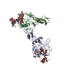
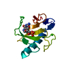
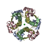
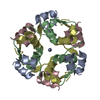
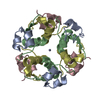
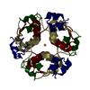
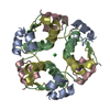
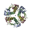
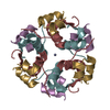
| Title | Crystal structure of humnan insulin Sr+2 complex | ||||||
 Components Components |
| ||||||
 Keywords Keywords |  HORMONE / Insulin / conformational state / metal binding / Carbohydrate metabolism / Cleavage on pair of basic residues / Diabetes mellitus / Disease mutation / Disulfide bond / Glucose metabolism / Secreted HORMONE / Insulin / conformational state / metal binding / Carbohydrate metabolism / Cleavage on pair of basic residues / Diabetes mellitus / Disease mutation / Disulfide bond / Glucose metabolism / Secreted | ||||||
| Function / homology |  Function and homology information Function and homology informationnegative regulation of NAD(P)H oxidase activity / negative regulation of glycogen catabolic process / regulation of cellular amino acid metabolic process / Signaling by Insulin receptor / IRS activation / nitric oxide-cGMP-mediated signaling / negative regulation of fatty acid metabolic process / Insulin processing / negative regulation of feeding behavior / regulation of protein secretion ...negative regulation of NAD(P)H oxidase activity / negative regulation of glycogen catabolic process / regulation of cellular amino acid metabolic process / Signaling by Insulin receptor / IRS activation / nitric oxide-cGMP-mediated signaling / negative regulation of fatty acid metabolic process / Insulin processing / negative regulation of feeding behavior / regulation of protein secretion / positive regulation of peptide hormone secretion / Regulation of gene expression in beta cells / positive regulation of respiratory burst / positive regulation of dendritic spine maintenance / alpha-beta T cell activation / negative regulation of acute inflammatory response / negative regulation of respiratory burst involved in inflammatory response / negative regulation of protein secretion / fatty acid homeostasis / Synthesis, secretion, and deacylation of Ghrelin / positive regulation of glycogen biosynthetic process / positive regulation of lipid biosynthetic process / Signal attenuation / FOXO-mediated transcription of oxidative stress, metabolic and neuronal genes / negative regulation of gluconeogenesis / positive regulation of nitric oxide mediated signal transduction / regulation of protein localization to plasma membrane / COPI-mediated anterograde transport / negative regulation of lipid catabolic process / negative regulation of oxidative stress-induced intrinsic apoptotic signaling pathway / negative regulation of reactive oxygen species biosynthetic process / positive regulation of insulin receptor signaling pathway / transport vesicle / positive regulation of protein autophosphorylation / Insulin receptor recycling / insulin-like growth factor receptor binding / NPAS4 regulates expression of target genes / positive regulation of protein metabolic process / neuron projection maintenance / endoplasmic reticulum-Golgi intermediate compartment membrane / positive regulation of brown fat cell differentiation / activation of protein kinase B activity / positive regulation of glycolytic process / Insulin receptor signalling cascade / positive regulation of mitotic nuclear division / Regulation of insulin secretion / positive regulation of nitric-oxide synthase activity / positive regulation of long-term synaptic potentiation / endosome lumen / positive regulation of cytokine production / acute-phase response / positive regulation of protein secretion / regulation of transmembrane transporter activity / positive regulation of cell differentiation / positive regulation of glucose import / negative regulation of proteolysis / regulation of synaptic plasticity / wound healing / insulin receptor binding / negative regulation of protein catabolic process / positive regulation of neuron projection development / hormone activity / cognition / Golgi lumen / vasodilation / positive regulation of protein localization to nucleus / glucose metabolic process / regulation of protein localization / glucose homeostasis / cell-cell signaling / insulin receptor signaling pathway / positive regulation of NF-kappaB transcription factor activity / PI5P, PP2A and IER3 Regulate PI3K/AKT Signaling / positive regulation of cell growth / secretory granule lumen / protease binding / positive regulation of MAPK cascade / positive regulation of phosphatidylinositol 3-kinase/protein kinase B signal transduction / positive regulation of cell migration / G protein-coupled receptor signaling pathway / Amyloid fiber formation / endoplasmic reticulum lumen / Golgi membrane / negative regulation of gene expression / positive regulation of cell population proliferation / positive regulation of gene expression / regulation of DNA-templated transcription / extracellular space / extracellular region / identical protein bindingSimilarity search - Function | ||||||
| Biological species |  Homo sapiens (human) Homo sapiens (human) | ||||||
| Method | X-RAY DIFFRACTION / MOLECULAR REPLACEMENT / Resolution: 1.9 Å | ||||||
 Authors Authors | Raghavendra, N. / Pattabhi, V. / Rajan, S.S. | ||||||
 Citation Citation | Journal: To be Published Title: Metal induced conformational changes in human insulin: Crystal structures of Sr+2, Ni+2 and Cu+2 complexes of human insulin Authors: Raghavendra, N. / Pattabhi, V. / Rajan, S.S. | ||||||
| History |
|
- Structure visualization
| Structure viewer | Molecule: MolmilJmol/JSmol |
|---|
- Downloads & links
-Download
| PDBx/mmCIF format | 3ilg.cif.gz | 33.1 KB | Display | PDBx/mmCIF format |
|---|---|---|---|---|
| PDB format | pdb3ilg.ent.gz | 23.2 KB | Display | PDB format |
| PDBx/mmJSON format | 3ilg.json.gz | Tree view | PDBx/mmJSON format | |
| Others |  Other downloads Other downloads |
-Validation report
| Arichive directory | https://data.pdbj.org/pub/pdb/validation_reports/il/3ilgftp://data.pdbj.org/pub/pdb/validation_reports/il/3ilg | HTTPS FTP |
|---|
-Related structure data
| Related structure data |  3incC 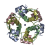 1msoS S: Starting model for refinement C: citing same article ( |
|---|---|
| Similar structure data |


-Links
 PDBj
PDBj


- Assembly
| Deposited unit | 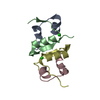
| |||||||||||||||||||||
|---|---|---|---|---|---|---|---|---|---|---|---|---|---|---|---|---|---|---|---|---|---|---|
| 1 | 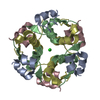
| |||||||||||||||||||||
| Unit cell |
| |||||||||||||||||||||
| Components on special symmetry positions |
|
-Components
| #1: Protein/peptide | Mass: 2383.698 Da / Num. of mol.: 2 / Source method: isolated from a natural source / Source: (natural) Homo sapiens (human) / References: UniProt: P01308#2: Protein/peptide | Mass: 3433.953 Da / Num. of mol.: 2 / Source method: isolated from a natural source / Source: (natural) Homo sapiens (human) / References: UniProt: P01308#3: Chemical | Strontium  Mass: 87.620 Da / Num. of mol.: 2 / Source method: obtained synthetically / Formula: Sr Mass: 87.620 Da / Num. of mol.: 2 / Source method: obtained synthetically / Formula: Sr#4: Water | ChemComp-HOH / | Water Mass: 18.015 Da / Num. of mol.: 57 / Source method: isolated from a natural source / Formula: H2O Mass: 18.015 Da / Num. of mol.: 57 / Source method: isolated from a natural source / Formula: H2O |
|---|
-Experimental details
-Experiment
| Experiment | Method: X-RAY DIFFRACTION / Number of used crystals: 1 |
|---|
- Sample preparation
| Crystal | Density Matthews: 1.84 Å3/Da / Density % sol: 33.29 % |
|---|---|
| Crystal grow | Temperature: 293 K / Method: vapor diffusion, hanging drop / pH: 6.8 Details: 0.1M Sodium Citrate, 1M Ammonium Sulphate, 0.1M Strontium Chloride, pH 6.8, VAPOR DIFFUSION, HANGING DROP, temperature 293.0K |
-Data collection
| Diffraction | Mean temperature: 100 K |
|---|---|
| Diffraction source | Source: ROTATING ANODE / Type: BRUKER AXS MICROSTAR / Wavelength: 1.54 Å |
| Detector | Type: MAR scanner 345 mm plate / Detector: IMAGE PLATE / Date: Dec 10, 2008 / Details: Mirror |
| Radiation | Protocol: SINGLE WAVELENGTH / Monochromatic (M) / Laue (L): M / Scattering type: x-ray |
| Radiation wavelength | Wavelength: 1.54 Å / Relative weight: 1 |
| Reflection | Resolution: 1.9→30 Å / Num. all: 6573 / Num. obs: 6243 / % possible obs: 99 % / Redundancy: 5.3 % / Biso Wilson estimate: 36.08 Å2 / Rmerge(I) obs: 0.0672 / Rsym value: 0.0573 / Net I/σ(I): 8.1 |
| Reflection shell | Resolution: 1.9→1.97 Å / Redundancy: 5.2 % / Rmerge(I) obs: 0.1745 / Mean I/σ(I) obs: 2.5 / Num. unique all: 6573 / Rsym value: 0.1643 / % possible all: 100 |
- Processing
| Software |
| ||||||||||||||||||||||||||||||||||||||||||||||||||||||||||||||||||||||||||||||||||||||||||||||||||||
|---|---|---|---|---|---|---|---|---|---|---|---|---|---|---|---|---|---|---|---|---|---|---|---|---|---|---|---|---|---|---|---|---|---|---|---|---|---|---|---|---|---|---|---|---|---|---|---|---|---|---|---|---|---|---|---|---|---|---|---|---|---|---|---|---|---|---|---|---|---|---|---|---|---|---|---|---|---|---|---|---|---|---|---|---|---|---|---|---|---|---|---|---|---|---|---|---|---|---|---|---|---|
| Refinement | Method to determine structure: MOLECULAR REPLACEMENT Starting model: 1MSO Resolution: 1.9→16.9 Å / Cor.coef. Fo:Fc: 0.948 / Cor.coef. Fo:Fc free: 0.883 / SU B: 4.222 / SU ML: 0.127 / Cross valid method: THROUGHOUT / ESU R: 0.208 / ESU R Free: 0.201 / Stereochemistry target values: MAXIMUM LIKELIHOOD / Details: HYDROGENS HAVE BEEN ADDED IN THE RIDING POSITIONS
| ||||||||||||||||||||||||||||||||||||||||||||||||||||||||||||||||||||||||||||||||||||||||||||||||||||
| Solvent computation | Ion probe radii: 0.8 Å / Shrinkage radii: 0.8 Å / VDW probe radii: 1.4 Å / Solvent model: MASK | ||||||||||||||||||||||||||||||||||||||||||||||||||||||||||||||||||||||||||||||||||||||||||||||||||||
| Displacement parameters | Biso mean: 25.11 Å2
| ||||||||||||||||||||||||||||||||||||||||||||||||||||||||||||||||||||||||||||||||||||||||||||||||||||
| Refinement step | Cycle: LAST / Resolution: 1.9→16.9 Å
| ||||||||||||||||||||||||||||||||||||||||||||||||||||||||||||||||||||||||||||||||||||||||||||||||||||
| Refine LS restraints |
| ||||||||||||||||||||||||||||||||||||||||||||||||||||||||||||||||||||||||||||||||||||||||||||||||||||
| LS refinement shell | Resolution: 1.9→1.949 Å / Rfactor Rfree error: 0.27804 / Total num. of bins used: 20
|
