 Movie
Movie Controller

[English] 日本語
 Yorodumi
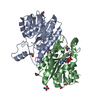
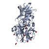
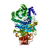
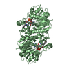
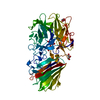
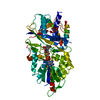
- PDB-3iii: 1.95 Angstrom Crystal Structure of CocE/NonD family hydrolase (SA... -
+ Open data

- Basic information
| Entry | Database: PDB / ID: 3iii | ||||||
|---|---|---|---|---|---|---|---|
| Title | 1.95 Angstrom Crystal Structure of CocE/NonD family hydrolase (SACOL2612) from Staphylococcus aureus | ||||||
 Components Components | CocE/NonD family hydrolase | ||||||
 Keywords Keywords |  HYDROLASE / structural genomics / Center for Structural Genomics of Infectious Diseases / CSGID HYDROLASE / structural genomics / Center for Structural Genomics of Infectious Diseases / CSGID | ||||||
| Function / homology |  Function and homology information Function and homology information | ||||||
| Biological species |  Staphylococcus aureus subsp. aureus COL (bacteria) Staphylococcus aureus subsp. aureus COL (bacteria) | ||||||
| Method | X-RAY DIFFRACTION / SYNCHROTRON / SAD / Resolution: 1.95 Å | ||||||
 Authors Authors | Osinski, T. / Chruszcz, M. / Domagalski, M.J. / Cymborowski, M. / Shumilin, I.A. / Skarina, T. / Onopriyenko, O. / Zimmerman, M.D. / Savchenko, A. / Edwards, A. ...Osinski, T. / Chruszcz, M. / Domagalski, M.J. / Cymborowski, M. / Shumilin, I.A. / Skarina, T. / Onopriyenko, O. / Zimmerman, M.D. / Savchenko, A. / Edwards, A. / Anderson, W.F. / Minor, W. / Center for Structural Genomics of Infectious Diseases (CSGID) | ||||||
 Citation Citation | Journal: To be Published Title: 1.95 Angstrom Crystal Structure of CocE/NonD family hydrolase (SACOL2612) from Staphylococcus aureus Authors: Osinski, T. / Chruszcz, M. / Domagalski, M.J. / Cymborowski, M. / Shumilin, I.A. / Skarina, T. / Onopriyenko, O. / Zimmerman, M.D. / Savchenko, A. / Edwards, A. / Anderson, W.F. / Minor, W. ...Authors: Osinski, T. / Chruszcz, M. / Domagalski, M.J. / Cymborowski, M. / Shumilin, I.A. / Skarina, T. / Onopriyenko, O. / Zimmerman, M.D. / Savchenko, A. / Edwards, A. / Anderson, W.F. / Minor, W. / Center for Structural Genomics of Infectious Diseases (CSGID) | ||||||
| History |
|
- Structure visualization
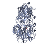
| Structure viewer | Molecule: MolmilJmol/JSmol |
|---|
- Downloads & links
-Download
| PDBx/mmCIF format | 3iii.cif.gz | 137.5 KB | Display | PDBx/mmCIF format |
|---|---|---|---|---|
| PDB format | pdb3iii.ent.gz | 111.6 KB | Display | PDB format |
| PDBx/mmJSON format | 3iii.json.gz | Tree view | PDBx/mmJSON format | |
| Others |  Other downloads Other downloads |
-Validation report
| Arichive directory | https://data.pdbj.org/pub/pdb/validation_reports/ii/3iiiftp://data.pdbj.org/pub/pdb/validation_reports/ii/3iii | HTTPS FTP |
|---|
-Related structure data
| Similar structure data | |
|---|---|
| Other databases |


-Links
 PDBj
PDBj


- Assembly
| Deposited unit | 
| ||||||||
|---|---|---|---|---|---|---|---|---|---|
| 1 |
| ||||||||
| Unit cell |
|
-Components
| #1: Protein | Mass: 64933.449 Da / Num. of mol.: 1 Source method: isolated from a genetically manipulated source Source: (gene. exp.) Staphylococcus aureus subsp. aureus COL (bacteria)Strain: Staphylococcus aureus subsp. aureus COL / Gene: SACOL2612 / Plasmid: pMCSG19c / Production host: Escherichia coli (E. coli) / Strain (production host): BL21-CodonPlus(DE3)-RIPL / References: UniProt: Q5HCV6, UniProt: A0A0H2WZL3*PLUS | ||||
|---|---|---|---|---|---|
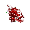
| #2: Chemical | ChemComp-NI / Nickel  Mass: 58.693 Da / Num. of mol.: 1 / Source method: obtained synthetically / Formula: Ni Mass: 58.693 Da / Num. of mol.: 1 / Source method: obtained synthetically / Formula: Ni | ||||
| #3: Chemical | ChemComp-CL / Chloride  Mass: 35.453 Da / Num. of mol.: 4 / Source method: obtained synthetically / Formula: Cl Mass: 35.453 Da / Num. of mol.: 4 / Source method: obtained synthetically / Formula: Cl#4: Chemical | ChemComp-PLM / | Palmitic acid  Mass: 256.424 Da / Num. of mol.: 1 / Source method: obtained synthetically / Formula: C16H32O2 Mass: 256.424 Da / Num. of mol.: 1 / Source method: obtained synthetically / Formula: C16H32O2#5: Water | ChemComp-HOH / | Water Mass: 18.015 Da / Num. of mol.: 556 / Source method: isolated from a natural source / Formula: H2O Mass: 18.015 Da / Num. of mol.: 556 / Source method: isolated from a natural source / Formula: H2O |
-Experimental details
-Experiment
| Experiment | Method: X-RAY DIFFRACTION / Number of used crystals: 1 |
|---|
- Sample preparation
| Crystal | Density Matthews: 2.25 Å3/Da / Density % sol: 45.35 % |
|---|---|
| Crystal grow | Temperature: 293 K / Method: vapor diffusion / pH: 7 Details: suberic acid, sebacic acid, hexadecanedioic acid, dodecanedioic acid 12mM each, PEG3350 20%, Hepes 0.1M pH 7.5, VAPOR DIFFUSION, temperature 293K |
-Data collection
| Diffraction | Mean temperature: 100 K |
|---|---|
| Diffraction source | Source: SYNCHROTRON / Site: APS  / Beamline: 19-BM / Wavelength: 0.9793 Å / Beamline: 19-BM / Wavelength: 0.9793 Å |
| Detector | Type: ADSC QUANTUM 210r / Detector: CCD / Date: Mar 18, 2009 / Details: mirrors |
| Radiation | Monochromator: Si 111 CHANNEL / Protocol: SINGLE WAVELENGTH / Monochromatic (M) / Laue (L): M / Scattering type: x-ray |
| Radiation wavelength | Wavelength: 0.9793 Å / Relative weight: 1 |
| Reflection | Resolution: 1.95→42.14 Å / Num. all: 44452 / Num. obs: 44452 / % possible obs: 100 % / Observed criterion σ(F): 0 / Observed criterion σ(I): -3 / Redundancy: 16.2 % / Rmerge(I) obs: 0.112 / Rsym value: 0.112 / Net I/σ(I): 29.1 |
| Reflection shell | Resolution: 1.95→1.98 Å / Redundancy: 15.3 % / Rmerge(I) obs: 0.637 / Mean I/σ(I) obs: 4.1 / Rsym value: 0.637 / % possible all: 100 |
- Processing
| Software |
| ||||||||||||||||||||||||||||||||||||||||||||||||||||||||||||||||||||||||||||||||||||||||||||||||||||||||||||||||||||||||||||||||||||||||||||||||||||||||||||||||||||||||||
|---|---|---|---|---|---|---|---|---|---|---|---|---|---|---|---|---|---|---|---|---|---|---|---|---|---|---|---|---|---|---|---|---|---|---|---|---|---|---|---|---|---|---|---|---|---|---|---|---|---|---|---|---|---|---|---|---|---|---|---|---|---|---|---|---|---|---|---|---|---|---|---|---|---|---|---|---|---|---|---|---|---|---|---|---|---|---|---|---|---|---|---|---|---|---|---|---|---|---|---|---|---|---|---|---|---|---|---|---|---|---|---|---|---|---|---|---|---|---|---|---|---|---|---|---|---|---|---|---|---|---|---|---|---|---|---|---|---|---|---|---|---|---|---|---|---|---|---|---|---|---|---|---|---|---|---|---|---|---|---|---|---|---|---|---|---|---|---|---|---|---|---|
| Refinement | Method to determine structure: SAD / Resolution: 1.95→42.14 Å / Cor.coef. Fo:Fc: 0.968 / Cor.coef. Fo:Fc free: 0.954 / SU B: 6.237 / SU ML: 0.082 / TLS residual ADP flag: LIKELY RESIDUAL / Cross valid method: THROUGHOUT / ESU R: 0.141 / ESU R Free: 0.127 Stereochemistry target values: MAXIMUM LIKELIHOOD WITH PHASES Details: HYDROGENS HAVE BEEN ADDED IN THE RIDING POSITIONS
| ||||||||||||||||||||||||||||||||||||||||||||||||||||||||||||||||||||||||||||||||||||||||||||||||||||||||||||||||||||||||||||||||||||||||||||||||||||||||||||||||||||||||||
| Solvent computation | Ion probe radii: 0.8 Å / Shrinkage radii: 0.8 Å / VDW probe radii: 1.2 Å / Solvent model: MASK | ||||||||||||||||||||||||||||||||||||||||||||||||||||||||||||||||||||||||||||||||||||||||||||||||||||||||||||||||||||||||||||||||||||||||||||||||||||||||||||||||||||||||||
| Displacement parameters | Biso mean: 8.694 Å2
| ||||||||||||||||||||||||||||||||||||||||||||||||||||||||||||||||||||||||||||||||||||||||||||||||||||||||||||||||||||||||||||||||||||||||||||||||||||||||||||||||||||||||||
| Refinement step | Cycle: LAST / Resolution: 1.95→42.14 Å
| ||||||||||||||||||||||||||||||||||||||||||||||||||||||||||||||||||||||||||||||||||||||||||||||||||||||||||||||||||||||||||||||||||||||||||||||||||||||||||||||||||||||||||
| Refine LS restraints |
| ||||||||||||||||||||||||||||||||||||||||||||||||||||||||||||||||||||||||||||||||||||||||||||||||||||||||||||||||||||||||||||||||||||||||||||||||||||||||||||||||||||||||||
| LS refinement shell | Resolution: 1.95→2.001 Å / Total num. of bins used: 20
| ||||||||||||||||||||||||||||||||||||||||||||||||||||||||||||||||||||||||||||||||||||||||||||||||||||||||||||||||||||||||||||||||||||||||||||||||||||||||||||||||||||||||||
| Refinement TLS params. | Method: refined / Refine-ID: X-RAY DIFFRACTION
| ||||||||||||||||||||||||||||||||||||||||||||||||||||||||||||||||||||||||||||||||||||||||||||||||||||||||||||||||||||||||||||||||||||||||||||||||||||||||||||||||||||||||||
| Refinement TLS group |
|
