 Movie
Movie Controller

[English] 日本語
 Yorodumi
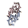
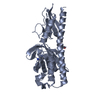
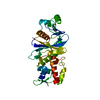
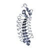
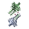
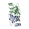
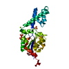
- PDB-3ht1: 1.2A structure of the polyketide cyclase RemF from Streptomyces r... -
+ Open data

- Basic information
| Entry | Database: PDB / ID: 3ht1 | ||||||
|---|---|---|---|---|---|---|---|
| Title | 1.2A structure of the polyketide cyclase RemF from Streptomyces resistomycificus | ||||||
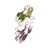
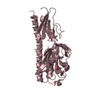
 Components Components | RemF protein | ||||||
 Keywords Keywords |  LYASE / cupin fold / ZN-binding / antibiotic biosynthesis / resistomycin / metalloprotein / cyclase LYASE / cupin fold / ZN-binding / antibiotic biosynthesis / resistomycin / metalloprotein / cyclase | ||||||
| Function / homology |  Function and homology information Function and homology information | ||||||
| Biological species |  Streptomyces resistomycificus (bacteria) Streptomyces resistomycificus (bacteria) | ||||||
| Method | X-RAY DIFFRACTION / SYNCHROTRON / SAD / Resolution: 1.2 Å | ||||||
 Authors Authors | Silvennoinen, L. / Sandalova, T. / Schneider, G. | ||||||
 Citation Citation | Journal: Febs Lett. / Year: 2009 Title: The polyketide cyclase RemF from Streptomyces resistomycificus contains an unusual octahedral zinc binding site Authors: Silvennoinen, L. / Sandalova, T. / Schneider, G. | ||||||
| History |
|
- Structure visualization
| Structure viewer | Molecule: MolmilJmol/JSmol |
|---|
- Downloads & links
-Download
| PDBx/mmCIF format | 3ht1.cif.gz | 76.9 KB | Display | PDBx/mmCIF format |
|---|---|---|---|---|
| PDB format | pdb3ht1.ent.gz | 56.4 KB | Display | PDB format |
| PDBx/mmJSON format | 3ht1.json.gz | Tree view | PDBx/mmJSON format | |
| Others |  Other downloads Other downloads |
-Validation report
| Arichive directory | https://data.pdbj.org/pub/pdb/validation_reports/ht/3ht1ftp://data.pdbj.org/pub/pdb/validation_reports/ht/3ht1 | HTTPS FTP |
|---|
-Related structure data


-Links
 PDBj
PDBj- Assembly
| Deposited unit | 
| ||||||||
|---|---|---|---|---|---|---|---|---|---|
| 1 | 
| ||||||||
| Unit cell |
|
-Components
| #1: Protein | Mass: 16383.433 Da / Num. of mol.: 1 Source method: isolated from a genetically manipulated source Source: (gene. exp.) Streptomyces resistomycificus (bacteria)Gene: remF / Plasmid: pRARE2 / Production host: Escherichia coli (E. coli) / Strain (production host): BL21-Gold (DE3) / References: UniProt: Q70DX5 |
|---|---|
| #2: Chemical | ChemComp-NI / Nickel  Mass: 58.693 Da / Num. of mol.: 1 / Source method: obtained synthetically / Formula: Ni Mass: 58.693 Da / Num. of mol.: 1 / Source method: obtained synthetically / Formula: Ni |
| #3: Water | ChemComp-HOH / Water Mass: 18.015 Da / Num. of mol.: 208 / Source method: isolated from a natural source / Formula: H2O Mass: 18.015 Da / Num. of mol.: 208 / Source method: isolated from a natural source / Formula: H2O |
-Experimental details
-Experiment
| Experiment | Method: X-RAY DIFFRACTION / Number of used crystals: 2 |
|---|
- Sample preparation
| Crystal | Density Matthews: 2.44 Å3/Da / Density % sol: 49.65 % |
|---|---|
| Crystal grow | Temperature: 293 K / Method: vapor diffusion, hanging drop / pH: 6.5 Details: 1M tri-sodium citrate, 0.1M sodium cacodylate, pH 6.5, VAPOR DIFFUSION, HANGING DROP, temperature 293K |
-Data collection
| Diffraction |
| |||||||||||||||
|---|---|---|---|---|---|---|---|---|---|---|---|---|---|---|---|---|
| Diffraction source |
| |||||||||||||||
| Detector |
| |||||||||||||||
| Radiation |
| |||||||||||||||
| Radiation wavelength |
| |||||||||||||||
| Reflection | Resolution: 1.15→47.9 Å / Num. all: 53620 / Num. obs: 53620 / % possible obs: 92.6 % / Observed criterion σ(F): 0 / Observed criterion σ(I): 0 / Redundancy: 7 % / Rmerge(I) obs: 0.063 / Net I/σ(I): 20.6 | |||||||||||||||
| Reflection shell | Resolution: 1.15→1.21 Å / Redundancy: 3.5 % / Mean I/σ(I) obs: 5.2 / Num. unique all: 5150 / Rsym value: 0.137 / % possible all: 68 |

- Processing
| Software |
| |||||||||||||||||||||||||||||||||
|---|---|---|---|---|---|---|---|---|---|---|---|---|---|---|---|---|---|---|---|---|---|---|---|---|---|---|---|---|---|---|---|---|---|---|
| Refinement | Method to determine structure: SAD / Resolution: 1.2→40 Å / Num. parameters: 12611 / Num. restraintsaints: 15950 / Cross valid method: FREE R / σ(F): 0 / σ(I): 0 / Stereochemistry target values: Engh & Huber Details: The structure was refined also with REFMAC 5.2.0019
| |||||||||||||||||||||||||||||||||
| Refine analyze | Num. disordered residues: 35 / Occupancy sum hydrogen: 1083 / Occupancy sum non hydrogen: 1345 | |||||||||||||||||||||||||||||||||
| Refinement step | Cycle: LAST / Resolution: 1.2→40 Å
| |||||||||||||||||||||||||||||||||
| Refine LS restraints |
|
