 Movie
Movie Controller

[English] 日本語
 Yorodumi
- PDB-3hmg: REFINEMENT OF THE INFLUENZA VIRUS HEMAGGLUTININ BY SIMULATED ANNEALING -
+ Open data

- Basic information
| Entry | Database: PDB / ID: 3hmg | |||||||||
|---|---|---|---|---|---|---|---|---|---|---|
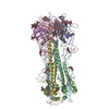
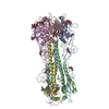
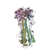
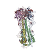
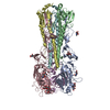
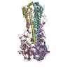
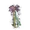
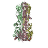
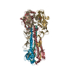
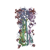
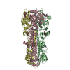
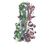
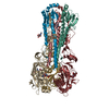
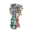
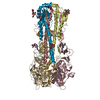
| Title | REFINEMENT OF THE INFLUENZA VIRUS HEMAGGLUTININ BY SIMULATED ANNEALING | |||||||||
 Components Components | (HEMAGGLUTININ ) x 2 ) x 2 | |||||||||
 Keywords Keywords | VIRAL PROTEIN / INFLUENZA VIRUS HEMAGGLUTININ | |||||||||
| Function / homology |  Function and homology informationviral budding from plasma membrane / clathrin-dependent endocytosis of virus by host cell / host cell surface receptor binding / fusion of virus membrane with host plasma membrane / fusion of virus membrane with host endosome membrane / viral envelope / virion attachment to host cell / host cell plasma membrane / virion membrane / membrane Function and homology informationviral budding from plasma membrane / clathrin-dependent endocytosis of virus by host cell / host cell surface receptor binding / fusion of virus membrane with host plasma membrane / fusion of virus membrane with host endosome membrane / viral envelope / virion attachment to host cell / host cell plasma membrane / virion membrane / membraneSimilarity search - Function | |||||||||
| Biological species |   Influenza A virus Influenza A virus | |||||||||
| Method | X-RAY DIFFRACTION / Resolution: 2.9 Å | |||||||||
 Authors Authors | Weis, W.I. / Bruenger, A.T. / Skehel, J.J. / Wiley, D.C. | |||||||||
 Citation Citation | Journal: J.Mol.Biol. / Year: 1990 Title: Refinement of the influenza virus hemagglutinin by simulated annealing. Authors: Weis, W.I. / Brunger, A.T. / Skehel, J.J. / Wiley, D.C. #1: Journal: Nature / Year: 1988Title: Structure of the Influenza Virus Haemagglutinin Complexes with its Receptor, Sialic Acid Authors: Weis, W. / Brown, J.H. / Cusack, S. / Paulson, J.C. / Skehel, J.J. / Wiley, D.C. #2: Journal: Nature / Year: 1984Title: Three-Dimensional Structure of an Antigenic Mutant of the Influenza Virus Haemagglutinin Authors: Knossow, M. / Daniels, R.S. / Douglas, A.R. / Skehel, J.J. / Wiley, D.C. #3: Journal: Nature / Year: 1981Title: Structure of the Haemagglutinin Membrane Glycoprotein of Influenza Virus at 3 Angstroms Resolution Authors: Wilson, I.A. / Skehel, J.J. / Wiley, D.C. #4: Journal: Nature / Year: 1981Title: Structural Identification of the Antibody-Binding Sites of Hong Kong Influenza Haemagglutinin and Their Involvement in Antigenic Variation Authors: Wiley, D.C. / Wilson, I.A. / Skehel, J.J. #5: Journal: J.Mol.Biol. / Year: 1977Title: Crystallization and X-Ray Diffraction Studies on the Haemagglutinin Glycoprotein from the Membrane of Influenza Virus Authors: Wiley, D.C. / Skehel, J.J. | |||||||||
| History |
|
- Structure visualization
| Structure viewer | Molecule: MolmilJmol/JSmol |
|---|
- Downloads & links
-Download
| PDBx/mmCIF format | 3hmg.cif.gz | 298 KB | Display | PDBx/mmCIF format |
|---|---|---|---|---|
| PDB format | pdb3hmg.ent.gz | 248.3 KB | Display | PDB format |
| PDBx/mmJSON format | 3hmg.json.gz | Tree view | PDBx/mmJSON format | |
| Others |  Other downloads Other downloads |
-Validation report
| Arichive directory | https://data.pdbj.org/pub/pdb/validation_reports/hm/3hmgftp://data.pdbj.org/pub/pdb/validation_reports/hm/3hmg | HTTPS FTP |
|---|
-Related structure data


-Links
 PDBj
PDBj


- Assembly
| Deposited unit | 
| ||||||||||||
|---|---|---|---|---|---|---|---|---|---|---|---|---|---|
| 1 |
| ||||||||||||
| Unit cell |
| ||||||||||||
| Atom site foot note | 1: RESIDUES PRO A 55, PRO C 55, PRO E 55 ARE CIS-PROLINES. 2: A TEMPERATURE FACTOR OF 65.0 OR LARGER SIGNIFIES AN ATOM WHOSE COORDINATES WERE GENERATED USING STEREOCHEMICAL CRITERIA. | ||||||||||||
| Noncrystallographic symmetry (NCS) | NCS oper:
| ||||||||||||
| Details | THERE IS ONE TRIMERIC HEMAGGLUTININ MOLECULE IN THE ASYMMETRIC UNIT, WITH THE MONOMERS RELATED TO EACH OTHER BY A NON-CRYSTALLOGRAPHIC THREE-FOLD AXIS. EACH MONOMER OF HEMAGGLUTININ CONSISTS OF TWO CHAINS, IDENTIFIED AS HA1 AND HA2 BY THE DEPOSITORS. CHAINS HA1 AND HA2 OF MONOMER 1 HAVE BEEN ASSIGNED CHAIN IDENTIFIERS *A* AND *B*, RESPECTIVELY. CHAINS HA1 AND HA2 OF MONOMER 2 HAVE BEEN ASSIGNED CHAIN IDENTIFIERS *C* AND *D*, RESPECTIVELY. CHAINS HA1 AND HA2 OF MONOMER 3 HAVE BEEN ASSIGNED CHAIN IDENTIFIERS *E* AND *F*, RESPECTIVELY. IN THE VIRUS, CHAIN HA1 CONSISTS OF 328 RESIDUES AND CHAIN HA2 CONSISTS OF 220 RESIDUES. HEMAGGLUTININ MAY BE SOLUBILIZED FROM THE VIRAL MEMBRANE BY BROMELAIN DIGESTION, WHICH REMOVES THE C-TERMINAL HYDROPHOBIC (ANCHORING) DOMAIN FROM CHAIN HA2. AFTER BROMELAIN DIGESTION CHAIN HA2 CONSISTS OF 175 RESIDUES, AS PRESENTED IN THIS ENTRY. IN THIS ENTRY RESIDUE LEU 226 IN CHAINS *A*, *C*, AND *E* HAS BEEN REPLACED BY GLN. THE WATERS ASSOCIATED WITH EACH MONOMER ARE PRESENTED IMMEDIATELY FOLLOWING CHAIN HA2 OF THAT MONOMER AND HAVE BEEN ASSIGNED THE SAME CHAIN INDICATOR (*B*, *D*, AND *F*). THE *MTRIX 1* TRANSFORMATION BELOW YIELDS APPROXIMATE COORDINATES FOR CHAINS *A* AND *B* WHEN APPLIED TO CHAINS *C* AND *D*. THE *MTRIX 2* TRANSFORMATION BELOW YIELDS APPROXIMATE COORDINATES FOR CHAINS *A* AND *B* WHEN APPLIED TO CHAINS *E* AND *F*. FOR THE RESTRAINED REGIONS, MONOMERS 1 AND 2 SUPERIMPOSE WITH AN RMS DEVIATION OF 0.030 ANGSTROMS AND MONOMERS 1 AND 3 SUPERIMPOSE WITH AN RMS DEVIATION OF 0.029 ANGSTROMS. |
-Components
| #1: Protein | Mass: 36080.430 Da / Num. of mol.: 3 Source method: isolated from a genetically manipulated source Source: (gene. exp.) Influenza A virus / Genus: Influenzavirus A / Strain: (A/Aichi/2/1968(H3N2)) / References: UniProt: P03437#2: Protein | Mass: 20212.350 Da / Num. of mol.: 3 Source method: isolated from a genetically manipulated source Source: (gene. exp.) Influenza A virus / Genus: Influenzavirus A / Strain: (A/Aichi/2/1968(H3N2)) / References: UniProt: P03437#3: Polysaccharide | / Mass: 586.542 Da / Num. of mol.: 3Source method: isolated from a genetically manipulated source #4: Sugar | ChemComp-NAG / N-Acetylglucosamine  Type: D-saccharide, beta linking / Mass: 221.208 Da / Num. of mol.: 12 Type: D-saccharide, beta linking / Mass: 221.208 Da / Num. of mol.: 12Source method: isolated from a genetically manipulated source Formula: C8H15NO6 #5: Water | ChemComp-HOH / | Water Mass: 18.015 Da / Num. of mol.: 33 / Source method: isolated from a natural source / Formula: H2O Mass: 18.015 Da / Num. of mol.: 33 / Source method: isolated from a natural source / Formula: H2O |
|---|
-Experimental details
-Experiment
| Experiment | Method: X-RAY DIFFRACTION |
|---|
- Sample preparation
| Crystal grow | *PLUS Method: microdialysisDetails: referred to 'Weis, W.', (1988) Nature, 333, 426-431 | ||||||||||||||||||||||||
|---|---|---|---|---|---|---|---|---|---|---|---|---|---|---|---|---|---|---|---|---|---|---|---|---|---|
| Components of the solutions | *PLUS
|
-Data collection
| Reflection | *PLUS Highest resolution: 2.9 Å / Lowest resolution: 12 Å / Num. obs: 74683 / Observed criterion σ(I): 3 / Rmerge(I) obs: 0.126 |
|---|
- Processing
| Software | Name: X-PLOR / Classification: refinement | ||||||||||||||||||||||||||||||||||||||||||||||||||||||||||||
|---|---|---|---|---|---|---|---|---|---|---|---|---|---|---|---|---|---|---|---|---|---|---|---|---|---|---|---|---|---|---|---|---|---|---|---|---|---|---|---|---|---|---|---|---|---|---|---|---|---|---|---|---|---|---|---|---|---|---|---|---|---|
| Refinement | Rfactor Rwork: 0.238 / Highest resolution: 2.9 Å Details: A TEMPERATURE FACTOR OF 65.0 OR LARGER SIGNIFIES AN ATOM WHOSE COORDINATES WERE GENERATED USING STEREOCHEMICAL CRITERIA. | ||||||||||||||||||||||||||||||||||||||||||||||||||||||||||||
| Refinement step | Cycle: LAST / Highest resolution: 2.9 Å
| ||||||||||||||||||||||||||||||||||||||||||||||||||||||||||||
| Refine LS restraints |
| ||||||||||||||||||||||||||||||||||||||||||||||||||||||||||||
| Refinement | *PLUS Highest resolution: 2.9 Å / Rfactor all: 0.238 | ||||||||||||||||||||||||||||||||||||||||||||||||||||||||||||
| Solvent computation | *PLUS | ||||||||||||||||||||||||||||||||||||||||||||||||||||||||||||
| Displacement parameters | *PLUS | ||||||||||||||||||||||||||||||||||||||||||||||||||||||||||||
| Refine LS restraints | *PLUS
|
