 Movie
Movie Controller

[English] 日本語
 Yorodumi
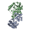
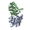
- PDB-3hdh: PIG HEART SHORT CHAIN L-3-HYDROXYACYL COA DEHYDROGENASE REVISITED... -
+ Open data

- Basic information
| Entry | Database: PDB / ID: 3hdh | ||||||
|---|---|---|---|---|---|---|---|
| Title | PIG HEART SHORT CHAIN L-3-HYDROXYACYL COA DEHYDROGENASE REVISITED: SEQUENCE ANALYSIS AND CRYSTAL STRUCTURE DETERMINATION | ||||||
 Components Components | PROTEIN (L-3-HYDROXYACYL COA DEHYDROGENASE) | ||||||
 Keywords Keywords |  OXIDOREDUCTASE / BETA OXIDATION / SCHAD / CATALYTIC ACTIVITY: L-3-HYDROXYACYL-COA + NAD(+) = 3-OXOACYL-COA + NADH OXIDOREDUCTASE / BETA OXIDATION / SCHAD / CATALYTIC ACTIVITY: L-3-HYDROXYACYL-COA + NAD(+) = 3-OXOACYL-COA + NADH | ||||||
| Function / homology |  Function and homology information Function and homology informationBeta oxidation of lauroyl-CoA to decanoyl-CoA-CoA / Beta oxidation of hexanoyl-CoA to butanoyl-CoA / Beta oxidation of butanoyl-CoA to acetyl-CoA / Beta oxidation of decanoyl-CoA to octanoyl-CoA-CoA / Beta oxidation of octanoyl-CoA to hexanoyl-CoA / 3-hydroxyacyl-CoA dehydrogenase / 3-hydroxyacyl-CoA dehydrogenase activity / fatty acid beta-oxidation / regulation of insulin secretion / NAD+ binding ...Beta oxidation of lauroyl-CoA to decanoyl-CoA-CoA / Beta oxidation of hexanoyl-CoA to butanoyl-CoA / Beta oxidation of butanoyl-CoA to acetyl-CoA / Beta oxidation of decanoyl-CoA to octanoyl-CoA-CoA / Beta oxidation of octanoyl-CoA to hexanoyl-CoA / 3-hydroxyacyl-CoA dehydrogenase / 3-hydroxyacyl-CoA dehydrogenase activity / fatty acid beta-oxidation / regulation of insulin secretion / NAD+ binding / positive regulation of cold-induced thermogenesis / mitochondrial matrix / nucleoplasm / identical protein bindingSimilarity search - Function | ||||||
| Biological species |  Sus scrofa (pig) Sus scrofa (pig) | ||||||
| Method | X-RAY DIFFRACTION / MOLECULAR REPLACEMENT / Resolution: 2.8 Å | ||||||
 Authors Authors | Barycki, J.J. / O'Brien, L.K. / Birktoft, J.J. / Strauss, A.W. / Banaszak, L.J. | ||||||
 Citation Citation | Journal: Protein Sci. / Year: 1999 Title: Pig heart short chain L-3-hydroxyacyl-CoA dehydrogenase revisited: sequence analysis and crystal structure determination. Authors: Barycki, J.J. / O'Brien, L.K. / Birktoft, J.J. / Strauss, A.W. / Banaszak, L.J. #1: Journal: Proc.Natl.Acad.Sci.USA / Year: 1987Title: Structure of L-3-Hydroxyacyl-Coenzyme a Dehydrogenase: Preliminary Chain Tracing at 2.8-A Resolution Authors: Birktoft, J.J. / Holden, H.M. / Hamlin, R. / Xuong, N.H. / Banaszak, L.J. | ||||||
| History |
|
- Structure visualization
| Structure viewer | Molecule: MolmilJmol/JSmol |
|---|
- Downloads & links
-Download
| PDBx/mmCIF format | 3hdh.cif.gz | 167.8 KB | Display | PDBx/mmCIF format |
|---|---|---|---|---|
| PDB format | pdb3hdh.ent.gz | 139.3 KB | Display | PDB format |
| PDBx/mmJSON format | 3hdh.json.gz | Tree view | PDBx/mmJSON format | |
| Others |  Other downloads Other downloads |
-Validation report
| Arichive directory | https://data.pdbj.org/pub/pdb/validation_reports/hd/3hdhftp://data.pdbj.org/pub/pdb/validation_reports/hd/3hdh | HTTPS FTP |
|---|
-Related structure data
| Related structure data | 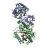 3hadS S: Starting model for refinement |
|---|---|
| Similar structure data |


-Links
 PDBj
PDBj


- Assembly
| Deposited unit | 
| ||||||||
|---|---|---|---|---|---|---|---|---|---|
| 1 | 
| ||||||||
| 2 | 
| ||||||||
| Unit cell |
| ||||||||
| Details | UNUSUAL SITUATION OF 3 SUBUNITS CONTAINED WITHIN THE ASYMMETRIC UNIT FOR A DIMERIC ENZYME. |
-Components
| #1: Protein | Mass: 32831.719 Da / Num. of mol.: 3 / Source method: isolated from a natural source / Source: (natural) Sus scrofa (pig) / Organ: HEART / Organelle: MITOCHONDRIALMitochondrionReferences: UniProt: P00348, 3-hydroxyacyl-CoA dehydrogenase#2: Chemical | Nicotinamide adenine dinucleotide  Mass: 663.425 Da / Num. of mol.: 3 / Source method: obtained synthetically / Formula: C21H27N7O14P2 / Comment: NAD*YM Mass: 663.425 Da / Num. of mol.: 3 / Source method: obtained synthetically / Formula: C21H27N7O14P2 / Comment: NAD*YM#3: Water | ChemComp-HOH / | Water Mass: 18.015 Da / Num. of mol.: 23 / Source method: isolated from a natural source / Formula: H2O Mass: 18.015 Da / Num. of mol.: 23 / Source method: isolated from a natural source / Formula: H2O |
|---|
-Experimental details
-Experiment
| Experiment | Method: X-RAY DIFFRACTION |
|---|
- Sample preparation
| Crystal | Density Matthews: 2.94 Å3/Da / Density % sol: 58 % / Description: USED DIMER AS A PROBE. | ||||||||||||||||||||||||||||||||||||
|---|---|---|---|---|---|---|---|---|---|---|---|---|---|---|---|---|---|---|---|---|---|---|---|---|---|---|---|---|---|---|---|---|---|---|---|---|---|
| Crystal grow | pH: 8 / Details: 10 MM TRIS, PH 8.0, + 1MM DTT + 11-14% PEG 6K | ||||||||||||||||||||||||||||||||||||
| Crystal grow | *PLUS Method: gel filtration | ||||||||||||||||||||||||||||||||||||
| Components of the solutions | *PLUS
|
-Data collection
| Diffraction | Mean temperature: 295 K |
|---|---|
| Diffraction source | Wavelength: 1.54 |
| Detector | Date: Jan 1, 1985 |
| Radiation | Protocol: SINGLE WAVELENGTH / Monochromatic (M) / Laue (L): M / Scattering type: x-ray |
| Radiation wavelength | Wavelength: 1.54 Å / Relative weight: 1 |
| Reflection | Resolution: 2.8→20 Å / Num. obs: 28840 / % possible obs: 98 % / Observed criterion σ(I): 0 / Redundancy: 4 % / Biso Wilson estimate: 58.7 Å2 / Rsym value: 0.046 / Net I/σ(I): 4.9 |
| Reflection shell | Resolution: 2.8→2.97 Å / % possible all: 85 |
| Reflection | *PLUS % possible obs: 98 % / Rmerge(I) obs: 0.046 |
- Processing
| Software |
| ||||||||||||||||||||||||||||||||||||||||||||||||||||||||||||||||||||||||||||||||
|---|---|---|---|---|---|---|---|---|---|---|---|---|---|---|---|---|---|---|---|---|---|---|---|---|---|---|---|---|---|---|---|---|---|---|---|---|---|---|---|---|---|---|---|---|---|---|---|---|---|---|---|---|---|---|---|---|---|---|---|---|---|---|---|---|---|---|---|---|---|---|---|---|---|---|---|---|---|---|---|---|---|
| Refinement | Method to determine structure: MOLECULAR REPLACEMENT Starting model: PDB ENTRY 3HAD Resolution: 2.8→20 Å / Rfactor Rfree error: 0.008 / Data cutoff high rms absF: 6104754.86 / Isotropic thermal model: RESTRAINED / Cross valid method: THROUGHOUT / σ(F): 1 Details: BULK SOLVENT MODEL USED POOR ELECTRON DENSITY OBSERVED FOR THIRD SUBUNIT (SUBUNIT C). THREE BREAKS WERE OBSERVED IN THE DENSITY AND SEVERAL RESIDUES HAVE BEEN ASSIGNED OCCUPANCIES OF 0.5
| ||||||||||||||||||||||||||||||||||||||||||||||||||||||||||||||||||||||||||||||||
| Displacement parameters | Biso mean: 62.5 Å2
| ||||||||||||||||||||||||||||||||||||||||||||||||||||||||||||||||||||||||||||||||
| Refine analyze |
| ||||||||||||||||||||||||||||||||||||||||||||||||||||||||||||||||||||||||||||||||
| Refinement step | Cycle: LAST / Resolution: 2.8→20 Å
| ||||||||||||||||||||||||||||||||||||||||||||||||||||||||||||||||||||||||||||||||
| Refine LS restraints |
| ||||||||||||||||||||||||||||||||||||||||||||||||||||||||||||||||||||||||||||||||
| LS refinement shell | Resolution: 2.8→2.97 Å / Rfactor Rfree error: 0.026 / Total num. of bins used: 6
| ||||||||||||||||||||||||||||||||||||||||||||||||||||||||||||||||||||||||||||||||
| Xplor file |
| ||||||||||||||||||||||||||||||||||||||||||||||||||||||||||||||||||||||||||||||||
| Software | *PLUS Name: CNS / Version: 0.3 / Classification: refinement | ||||||||||||||||||||||||||||||||||||||||||||||||||||||||||||||||||||||||||||||||
| Refinement | *PLUS σ(F): 1 / % reflection Rfree: 4.9 % / Rfactor obs: 0.223 | ||||||||||||||||||||||||||||||||||||||||||||||||||||||||||||||||||||||||||||||||
| Solvent computation | *PLUS | ||||||||||||||||||||||||||||||||||||||||||||||||||||||||||||||||||||||||||||||||
| Displacement parameters | *PLUS Biso mean: 62.5 Å2 | ||||||||||||||||||||||||||||||||||||||||||||||||||||||||||||||||||||||||||||||||
| Refine LS restraints | *PLUS
| ||||||||||||||||||||||||||||||||||||||||||||||||||||||||||||||||||||||||||||||||
| LS refinement shell | *PLUS Rfactor Rfree: 0.329 / % reflection Rfree: 5.3 % / Rfactor Rwork: 0.306 |
