 Movie
Movie Controller

[English] 日本語
 Yorodumi
- PDB-3gwh: Crystallographic Ab Initio protein solution far below atomic reso... -
+ Open data

- Basic information
| Entry | Database: PDB / ID: 3gwh | ||||||
|---|---|---|---|---|---|---|---|
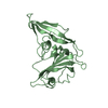
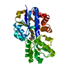
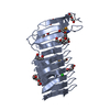
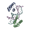
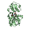
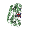
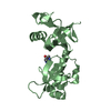
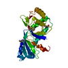
| Title | Crystallographic Ab Initio protein solution far below atomic resolution | ||||||
 Components Components | Transcriptional antiterminator (BglG family) | ||||||
 Keywords Keywords |  TRANSCRIPTION / extended helix bundle / Ab Initio / Structure solution / ARCIMBOLDO / PHASER / SHELXE TRANSCRIPTION / extended helix bundle / Ab Initio / Structure solution / ARCIMBOLDO / PHASER / SHELXE | ||||||
| Function / homology |  Function and homology information Function and homology information | ||||||
| Biological species |  Bacillus subtilis (bacteria) Bacillus subtilis (bacteria) | ||||||
| Method | X-RAY DIFFRACTION / AB INITIO / Resolution: 1.95 Å | ||||||
 Authors Authors | Rodriguez, D.D. / Grosse, C. / Himmel, S. / Gonzalez, C. / Becker, S. / Sheldrick, G.M. / Uson, I. | ||||||
 Citation Citation | Journal: Nat.Methods / Year: 2009 Title: Crystallographic ab initio protein structure solution below atomic resolution Authors: Rodriguez, D.D. / Grosse, C. / Himmel, S. / Gonzalez, C. / de Ilarduya, I.M. / Becker, S. / Sheldrick, G.M. / Uson, I. | ||||||
| History |
|
- Structure visualization
| Structure viewer | Molecule: MolmilJmol/JSmol |
|---|
- Downloads & links
-Download
| PDBx/mmCIF format | 3gwh.cif.gz | 53.7 KB | Display | PDBx/mmCIF format |
|---|---|---|---|---|
| PDB format | pdb3gwh.ent.gz | 39.4 KB | Display | PDB format |
| PDBx/mmJSON format | 3gwh.json.gz | Tree view | PDBx/mmJSON format | |
| Others |  Other downloads Other downloads |
-Validation report
| Arichive directory | https://data.pdbj.org/pub/pdb/validation_reports/gw/3gwhftp://data.pdbj.org/pub/pdb/validation_reports/gw/3gwh | HTTPS FTP |
|---|
-Related structure data
| Similar structure data |
|---|


-Links
 PDBj
PDBj- Assembly
| Deposited unit | 
| ||||||||
|---|---|---|---|---|---|---|---|---|---|
| 1 |
| ||||||||
| Unit cell |
|
-Components
| #1: Protein | Mass: 13154.480 Da / Num. of mol.: 2 / Fragment: UNP residues 178-285 Source method: isolated from a genetically manipulated source Details: GST fusion protein, pGEX2TEV derived from commercial pGEX2T Source: (gene. exp.) Bacillus subtilis (bacteria) / Strain: 168 / Gene: BSU13880, glcT / Plasmid: pGEX2TEV / Production host: Escherichia coli (E. coli) / Strain (production host): BL21 / References: UniProt: O31691#2: Chemical | Phosphate  Mass: 94.971 Da / Num. of mol.: 2 / Source method: obtained synthetically / Formula: PO4 Mass: 94.971 Da / Num. of mol.: 2 / Source method: obtained synthetically / Formula: PO4#3: Water | ChemComp-HOH / | Water Mass: 18.015 Da / Num. of mol.: 29 / Source method: isolated from a natural source / Formula: H2O Mass: 18.015 Da / Num. of mol.: 29 / Source method: isolated from a natural source / Formula: H2O |
|---|
-Experimental details
-Experiment
| Experiment | Method: X-RAY DIFFRACTION / Number of used crystals: 1 |
|---|
- Sample preparation
| Crystal | Density Matthews: 1.77 Å3/Da / Density % sol: 30.59 % |
|---|---|
| Crystal grow | Temperature: 293 K / Method: vapor diffusion, sitting drop / pH: 6 Details: 2.4M (NH4)2SO4, 0.1M citric acid, pH6.0, vapor diffusion, sitting drop, temperature 293K, VAPOR DIFFUSION, SITTING DROP |
-Data collection
| Diffraction | Mean temperature: 100 K |
|---|---|
| Diffraction source | Source: ROTATING ANODE / Type: MACSCIENCE / Wavelength: 1.5418 Å |
| Detector | Type: BRUKER SMART 6000 / Detector: CCD / Date: Aug 22, 2008 / Details: MULTI-LAYER INCOATEC OPTICS |
| Radiation | Monochromator: MULTI-LAYER INCOATEC OPTICS / Protocol: SINGLE WAVELENGTH / Monochromatic (M) / Laue (L): M / Scattering type: x-ray |
| Radiation wavelength | Wavelength: 1.5418 Å / Relative weight: 1 |
| Reflection | Resolution: 1.95→35.98 Å / Num. obs: 12855 / % possible obs: 99.6 % / Observed criterion σ(I): -3 / Redundancy: 16.7 % / Rmerge(I) obs: 0.0906 / Net I/σ(I): 20.21 |
| Reflection shell | Resolution: 1.95→2.04 Å / Redundancy: 1.75 % / Mean I/σ(I) obs: 2.45 / Num. unique all: 1786 / % possible all: 96.8 |
- Processing
| Software |
| |||||||||||||||||||||||||||||||||||||||||||||||||||||||||||||||||||||||||||||||||||||
|---|---|---|---|---|---|---|---|---|---|---|---|---|---|---|---|---|---|---|---|---|---|---|---|---|---|---|---|---|---|---|---|---|---|---|---|---|---|---|---|---|---|---|---|---|---|---|---|---|---|---|---|---|---|---|---|---|---|---|---|---|---|---|---|---|---|---|---|---|---|---|---|---|---|---|---|---|---|---|---|---|---|---|---|---|---|---|
| Refinement | Method to determine structure: AB INITIO / Resolution: 1.95→35.98 Å / Cor.coef. Fo:Fc: 0.949 / Cor.coef. Fo:Fc free: 0.94 / WRfactor Rfree: 0.229 / WRfactor Rwork: 0.185 / Occupancy max: 1 / Occupancy min: 0.5 / FOM work R set: 0.837 / SU B: 4.629 / SU ML: 0.133 / SU R Cruickshank DPI: 0.214 / SU Rfree: 0.177 / Cross valid method: THROUGHOUT / σ(F): 0 / σ(I): 2 / ESU R: 0.214 / ESU R Free: 0.177 / Stereochemistry target values: MAXIMUM LIKELIHOOD Details: HYDROGENS HAVE BEEN ADDED IN THE RIDING POSITIONS, The data were non-merohedrally twinned. The twinning fraction was 0.729/0.271, The data were detwinned with twinabs for refinement. The ...Details: HYDROGENS HAVE BEEN ADDED IN THE RIDING POSITIONS, The data were non-merohedrally twinned. The twinning fraction was 0.729/0.271, The data were detwinned with twinabs for refinement. The crystal was a two-component non-merohedral twin. The frames were indexed with CELL_NOW, integrated with SAINT and scaled with TWINABS (all programs from Bruker AXS) to obtain the merged intensities of the unique reflections from measurements of all 99947 reflections of domain 1, 98274 reflections of domain 2 and 25843 reflections for which both domains overlapped.
| |||||||||||||||||||||||||||||||||||||||||||||||||||||||||||||||||||||||||||||||||||||
| Solvent computation | Ion probe radii: 0.8 Å / Shrinkage radii: 0.8 Å / VDW probe radii: 1.4 Å / Solvent model: BABINET MODEL WITH MASK | |||||||||||||||||||||||||||||||||||||||||||||||||||||||||||||||||||||||||||||||||||||
| Displacement parameters | Biso max: 59.6 Å2 / Biso mean: 27.144 Å2 / Biso min: 8.68 Å2
| |||||||||||||||||||||||||||||||||||||||||||||||||||||||||||||||||||||||||||||||||||||
| Refinement step | Cycle: LAST / Resolution: 1.95→35.98 Å
| |||||||||||||||||||||||||||||||||||||||||||||||||||||||||||||||||||||||||||||||||||||
| Refine LS restraints |
| |||||||||||||||||||||||||||||||||||||||||||||||||||||||||||||||||||||||||||||||||||||
| LS refinement shell | Resolution: 1.945→1.996 Å / Total num. of bins used: 20
|
