 Movie
Movie Controller

[English] 日本語
 Yorodumi
- PDB-3gtu: LIGAND-FREE HETERODIMERIC HUMAN GLUTATHIONE S-TRANSFERASE M2-3 (E... -
+ Open data

- Basic information
| Entry | Database: PDB / ID: 3gtu | ||||||
|---|---|---|---|---|---|---|---|
| Title | LIGAND-FREE HETERODIMERIC HUMAN GLUTATHIONE S-TRANSFERASE M2-3 (EC 2.5.1.18), MONOCLINIC CRYSTAL FORM | ||||||
 Components Components | (GLUTATHIONE S-TRANSFERASE ) x 2 ) x 2 | ||||||
 Keywords Keywords | TRANSFERASE / GLUTATHIONE / CONJUGATION / DETOXIFICATION / CYTOSOLIC / HETERODIMER | ||||||
| Function / homology |  Function and homology information Function and homology informationestablishment of blood-nerve barrier / sperm fibrous sheath / regulation of skeletal muscle contraction by regulation of release of sequestered calcium ion / nitrobenzene metabolic process / cellular detoxification of nitrogen compound / hepoxilin biosynthetic process / glutathione binding / glutathione derivative biosynthetic process / linoleic acid metabolic process / Glutathione conjugation ...establishment of blood-nerve barrier / sperm fibrous sheath / regulation of skeletal muscle contraction by regulation of release of sequestered calcium ion / nitrobenzene metabolic process / cellular detoxification of nitrogen compound / hepoxilin biosynthetic process / glutathione binding / glutathione derivative biosynthetic process / linoleic acid metabolic process / Glutathione conjugation / glutathione peroxidase activity / relaxation of cardiac muscle / intercellular bridge / positive regulation of ryanodine-sensitive calcium-release channel activity / cellular response to caffeine / glutathione transferase / glutathione transferase activity / negative regulation of ryanodine-sensitive calcium-release channel activity / regulation of cardiac muscle contraction by regulation of the release of sequestered calcium ion / glutathione metabolic process / xenobiotic catabolic process / regulation of release of sequestered calcium ion into cytosol by sarcoplasmic reticulum / sarcoplasmic reticulum / fatty acid binding / response to estrogen / signaling receptor binding / enzyme binding / protein homodimerization activity / extracellular exosome / identical protein binding / nucleus / cytosol / cytoplasmSimilarity search - Function | ||||||
| Biological species |  Homo sapiens (human) Homo sapiens (human) | ||||||
| Method | X-RAY DIFFRACTION / MOLECULAR REPLACEMENT / Resolution: 2.8 Å | ||||||
 Authors Authors | Patskovsky, Y.V. / Patskovska, L.N. / Listowsky, I. | ||||||
 Citation Citation | Journal: Biochemistry / Year: 1999 Title: An asparagine-phenylalanine substitution accounts for catalytic differences between hGSTM3-3 and other human class mu glutathione S-transferases. Authors: Patskovsky, Y.V. / Patskovska, L.N. / Listowsky, I. #1: Journal: J.Mol.Biol. / Year: 1994Title: Crystal Structure of Human Class Mu Glutathione Transferase Gstm2-2. Effects of Lattice Packing on Conformational Heterogeneity Authors: Raghunathan, S. / Chandross, R.J. / Kretsinger, R.H. / Allison, T.J. / Penington, C.J. / Rule, G.S. #2: Journal: Proc.Natl.Acad.Sci.USA / Year: 1991Title: Cloning, Expression, and Characterization of a Class-Mu Glutathione Transferase from Human Muscle, the Product of the Gst4 Locus Authors: Vorachek, W.R. / Pearson, W.R. / Rule, G.S. #3: Journal: J.Biol.Chem. / Year: 1990Title: A Distinct Human Testis and Brain Mu-Class Glutathione S-Transferase. Molecular Cloning and Characterization of a Form Present Even in Individuals Lacking Hepatic Type Mu Isoenzymes Authors: Campbell, E. / Takahashi, Y. / Abramovitz, M. / Peretz, M. / Listowsky, I. | ||||||
| History |
|
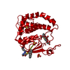
- Structure visualization
| Structure viewer | Molecule: MolmilJmol/JSmol |
|---|
- Downloads & links
-Download
| PDBx/mmCIF format | 3gtu.cif.gz | 184.8 KB | Display | PDBx/mmCIF format |
|---|---|---|---|---|
| PDB format | pdb3gtu.ent.gz | 151.8 KB | Display | PDB format |
| PDBx/mmJSON format | 3gtu.json.gz | Tree view | PDBx/mmJSON format | |
| Others |  Other downloads Other downloads |
-Validation report
| Arichive directory | https://data.pdbj.org/pub/pdb/validation_reports/gt/3gtuftp://data.pdbj.org/pub/pdb/validation_reports/gt/3gtu | HTTPS FTP |
|---|
-Related structure data
| Related structure data |  4gtuC 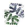 2gtuS S: Starting model for refinement C: citing same article ( |
|---|---|
| Similar structure data |


-Links
 PDBj
PDBj

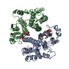
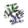
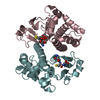
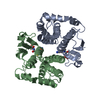
- Assembly
| Deposited unit | 
| ||||||||||||
|---|---|---|---|---|---|---|---|---|---|---|---|---|---|
| 1 | 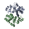
| ||||||||||||
| 2 | 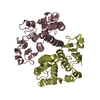
| ||||||||||||
| 3 | 
| ||||||||||||
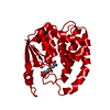
| Unit cell |
| ||||||||||||
| Noncrystallographic symmetry (NCS) | NCS oper:
|
-Components
| #1: Protein | Mass: 25645.457 Da / Num. of mol.: 2 Source method: isolated from a genetically manipulated source Details: LIGAND-FREE, HETERODIMER / Source: (gene. exp.) Homo sapiens (human)Description: THE GSTM2 AND GSTM3 CDNA WERE AMPLIFIED USING RT-PCR AND SUBCLONED INTO A PET3A COEXPRESSION VECTOR. SYNTHETIC GENE. Cell line: HELA / Cellular location: CYTOPLASM / Gene: GSTM2, GSTM3 / Plasmid: PET3A-GSTM2-3 / Species (production host): Escherichia coli / Gene (production host): GSTM2, GSTM3 / Production host:  Escherichia coli BL21(DE3) (bacteria) / Strain (production host): BL21 (DE3) / References: UniProt: P28161, glutathione transferase Escherichia coli BL21(DE3) (bacteria) / Strain (production host): BL21 (DE3) / References: UniProt: P28161, glutathione transferase#2: Protein | Mass: 26460.410 Da / Num. of mol.: 2 Source method: isolated from a genetically manipulated source Details: LIGAND-FREE, HETERODIMER / Source: (gene. exp.) Homo sapiens (human)Description: THE GSTM2 AND GSTM3 CDNA WERE AMPLIFIED USING RT-PCR AND SUBCLONED INTO A PET3A COEXPRESSION VECTOR. SYNTHETIC GENE. Cell line: HELA / Cellular location: CYTOPLASM / Gene: GSTM2, GSTM3 / Plasmid: PET3A-GSTM2-3 / Species (production host): Escherichia coli / Gene (production host): GSTM2, GSTM3 / Production host: Escherichia coli BL21(DE3) (bacteria) / Strain (production host): BL21 (DE3) / References: UniProt: P21266, glutathione transferase#3: Water | ChemComp-HOH / | Water Mass: 18.015 Da / Num. of mol.: 72 / Source method: isolated from a natural source / Formula: H2O Mass: 18.015 Da / Num. of mol.: 72 / Source method: isolated from a natural source / Formula: H2O |
|---|
-Experimental details
-Experiment
| Experiment | Method: X-RAY DIFFRACTION / Number of used crystals: 1 |
|---|
- Sample preparation
| Crystal | Density Matthews: 2.65 Å3/Da / Density % sol: 50 % |
|---|---|
| Crystal grow | pH: 7.5 / Details: pH 7.5 |
| Crystal | *PLUS |
| Crystal grow | *PLUS Method: unknown |
-Data collection
| Diffraction | Mean temperature: 289 K |
|---|---|
| Diffraction source | Source: ROTATING ANODE / Type: RIGAKU RUH2R / Wavelength: 1.5418 |
| Detector | Type: SIEMENS-NICOLET X100 / Detector: AREA DETECTOR / Date: Jul 1, 1997 |
| Radiation | Monochromator: GRAPHITE(002) / Monochromatic (M) / Laue (L): M / Scattering type: x-ray |
| Radiation wavelength | Wavelength: 1.5418 Å / Relative weight: 1 |
| Reflection | Resolution: 2.8→10 Å / Num. obs: 25387 / % possible obs: 94.41 % / Observed criterion σ(I): 2 / Redundancy: 2.4 % / Rmerge(I) obs: 0.113 / Net I/σ(I): 3.5 |
| Reflection shell | Resolution: 2.8→2.9 Å / Redundancy: 1.71 % / Rmerge(I) obs: 0.341 / % possible all: 69.4 |
- Processing
| Software |
| ||||||||||||||||||||||||||||||||||||||||||||||||||||||||||||||||||||||||||||||||
|---|---|---|---|---|---|---|---|---|---|---|---|---|---|---|---|---|---|---|---|---|---|---|---|---|---|---|---|---|---|---|---|---|---|---|---|---|---|---|---|---|---|---|---|---|---|---|---|---|---|---|---|---|---|---|---|---|---|---|---|---|---|---|---|---|---|---|---|---|---|---|---|---|---|---|---|---|---|---|---|---|---|
| Refinement | Method to determine structure: MOLECULAR REPLACEMENT Starting model: PDB ENTRY 2GTU Resolution: 2.8→8 Å / Rfactor Rfree error: 0.01 / Data cutoff high absF: 1000000 / Data cutoff low absF: 0.001 / Isotropic thermal model: RESTRAINED / Cross valid method: THROUGHOUT / σ(F): 2
| ||||||||||||||||||||||||||||||||||||||||||||||||||||||||||||||||||||||||||||||||
| Refine analyze |
| ||||||||||||||||||||||||||||||||||||||||||||||||||||||||||||||||||||||||||||||||
| Refinement step | Cycle: LAST / Resolution: 2.8→8 Å
| ||||||||||||||||||||||||||||||||||||||||||||||||||||||||||||||||||||||||||||||||
| Refine LS restraints |
| ||||||||||||||||||||||||||||||||||||||||||||||||||||||||||||||||||||||||||||||||
| LS refinement shell | Resolution: 2.8→2.92 Å / Rfactor Rfree error: 0.014 / Total num. of bins used: 8
| ||||||||||||||||||||||||||||||||||||||||||||||||||||||||||||||||||||||||||||||||
| Xplor file |
| ||||||||||||||||||||||||||||||||||||||||||||||||||||||||||||||||||||||||||||||||
| Software | *PLUS Name: X-PLOR / Version: 3.851 / Classification: refinement | ||||||||||||||||||||||||||||||||||||||||||||||||||||||||||||||||||||||||||||||||
| Refinement | *PLUS Rfactor Rfree: 0.27 | ||||||||||||||||||||||||||||||||||||||||||||||||||||||||||||||||||||||||||||||||
| Solvent computation | *PLUS | ||||||||||||||||||||||||||||||||||||||||||||||||||||||||||||||||||||||||||||||||
| Displacement parameters | *PLUS | ||||||||||||||||||||||||||||||||||||||||||||||||||||||||||||||||||||||||||||||||
| Refine LS restraints | *PLUS
|
