 Movie
Movie Controller


+ Open data

- Basic information
| Entry | Database: PDB / ID: 3gnf | ||||||
|---|---|---|---|---|---|---|---|
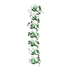
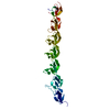
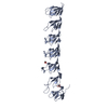
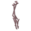
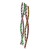
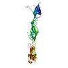
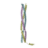
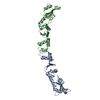
| Title | P1 Crystal structure of the N-terminal R1-R7 of murine MVP | ||||||
 Components Components | Major vault protein | ||||||
 Keywords Keywords | STRUCTURAL PROTEIN / beta sheets / Phosphoprotein / Ribonucleoprotein | ||||||
| Function / homology |  Function and homology information Function and homology informationprotein activation cascade / negative regulation of protein autophosphorylation / ERBB signaling pathway / negative regulation of epidermal growth factor receptor signaling pathway / Neutrophil degranulation / protein phosphatase binding / cell population proliferation / cytoskeleton / ribonucleoprotein complex / protein kinase binding ...protein activation cascade / negative regulation of protein autophosphorylation / ERBB signaling pathway / negative regulation of epidermal growth factor receptor signaling pathway / Neutrophil degranulation / protein phosphatase binding / cell population proliferation / cytoskeleton / ribonucleoprotein complex / protein kinase binding / perinuclear region of cytoplasm / identical protein binding / nucleus / cytosol / cytoplasmSimilarity search - Function | ||||||
| Biological species |  Mus musculus (house mouse) Mus musculus (house mouse) | ||||||
| Method | X-RAY DIFFRACTION / SYNCHROTRON / MOLECULAR REPLACEMENT / MAD / Resolution: 2.1 Å | ||||||
 Authors Authors | Querol-Audi, J. / Casanas, A. / Uson, I. / Luque, D. / Caston, J.R. / Fita, I. / Verdaguer, N. | ||||||
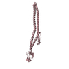
 Citation Citation | Journal: Embo J. / Year: 2009 Title: The mechanism of vault opening from the high resolution structure of the N-terminal repeats of MVP Authors: Querol-Audi, J. / Casanas, A. / Uson, I. / Luque, D. / Caston, J.R. / Fita, I. / Verdaguer, N. | ||||||
| History |
|
- Structure visualization
| Structure viewer | Molecule: MolmilJmol/JSmol |
|---|
- Downloads & links
-Download
| PDBx/mmCIF format | 3gnf.cif.gz | 86.8 KB | Display | PDBx/mmCIF format |
|---|---|---|---|---|
| PDB format | pdb3gnf.ent.gz | 64 KB | Display | PDB format |
| PDBx/mmJSON format | 3gnf.json.gz | Tree view | PDBx/mmJSON format | |
| Others |  Other downloads Other downloads |
-Validation report
| Arichive directory | https://data.pdbj.org/pub/pdb/validation_reports/gn/3gnfftp://data.pdbj.org/pub/pdb/validation_reports/gn/3gnf | HTTPS FTP |
|---|
-Related structure data
| Related structure data |  3gf5SC  3gngC S: Starting model for refinement C: citing same article ( |
|---|---|
| Similar structure data |

-Links
 PDBj
PDBj


- Assembly
| Deposited unit | 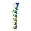
| ||||||||
|---|---|---|---|---|---|---|---|---|---|
| 1 |
| ||||||||
| Unit cell |
|
-Components
| #1: Protein | / MVP Mass: 43735.777 Da / Num. of mol.: 1 / Fragment: R1-R7 domain, UNP residues 1-383 Source method: isolated from a genetically manipulated source Source: (gene. exp.) Mus musculus (house mouse) / Gene: Mvp / Plasmid: pGex-6P1 / Production host:  Escherichia coli (E. coli) / Strain (production host): BL21(DE3) / References: UniProt: Q9EQK5 Escherichia coli (E. coli) / Strain (production host): BL21(DE3) / References: UniProt: Q9EQK5 |
|---|---|
| #2: Water | ChemComp-HOH / Water Mass: 18.015 Da / Num. of mol.: 59 / Source method: isolated from a natural source / Formula: H2O Mass: 18.015 Da / Num. of mol.: 59 / Source method: isolated from a natural source / Formula: H2O |
-Experimental details
-Experiment
| Experiment | Method: X-RAY DIFFRACTION / Number of used crystals: 1 |
|---|
- Sample preparation
| Crystal | Density Matthews: 2.68 Å3/Da / Density % sol: 54.12 % |
|---|---|
| Crystal grow | Temperature: 277 K / Method: vapor diffusion, hanging drop / pH: 5.6 Details: 20%PEG 5000, 0.1M Sodium Citrate, pH 5.6, VAPOR DIFFUSION, HANGING DROP, temperature 277K |
-Data collection
| Diffraction | Mean temperature: 100 K |
|---|---|
| Diffraction source | Source: SYNCHROTRON / Site: ESRF  / Beamline: ID23-1 / Wavelength: 0.979 Å / Beamline: ID23-1 / Wavelength: 0.979 Å |
| Detector | Type: ADSC QUANTUM 315r / Detector: CCD / Date: Apr 12, 2007 |
| Radiation | Monochromator: Si 111 CHANNEL / Protocol: SINGLE WAVELENGTH / Monochromatic (M) / Laue (L): M / Scattering type: x-ray |
| Radiation wavelength | Wavelength: 0.979 Å / Relative weight: 1 |
| Reflection | Resolution: 2.1→30 Å / Num. obs: 26128 / Observed criterion σ(F): 2 / Observed criterion σ(I): 1 / Rmerge(I) obs: 0.031 / Net I/σ(I): 14.3 |
-Phasing
| Phasing | Method: MAD |
|---|
- Processing
| Software |
| ||||||||||||||||||||||||||||||||||||||||||||||||||||||||||||||||||||||||||||||||||||||||||||||||||||||||||||||||||||||||||||||||||||||||||||||||||||||||||||||||||||||||||
|---|---|---|---|---|---|---|---|---|---|---|---|---|---|---|---|---|---|---|---|---|---|---|---|---|---|---|---|---|---|---|---|---|---|---|---|---|---|---|---|---|---|---|---|---|---|---|---|---|---|---|---|---|---|---|---|---|---|---|---|---|---|---|---|---|---|---|---|---|---|---|---|---|---|---|---|---|---|---|---|---|---|---|---|---|---|---|---|---|---|---|---|---|---|---|---|---|---|---|---|---|---|---|---|---|---|---|---|---|---|---|---|---|---|---|---|---|---|---|---|---|---|---|---|---|---|---|---|---|---|---|---|---|---|---|---|---|---|---|---|---|---|---|---|---|---|---|---|---|---|---|---|---|---|---|---|---|---|---|---|---|---|---|---|---|---|---|---|---|---|---|---|
| Refinement | Method to determine structure: MOLECULAR REPLACEMENT Starting model: PDB ENTRY 3GF5 Resolution: 2.1→30 Å / Cor.coef. Fo:Fc: 0.938 / Cor.coef. Fo:Fc free: 0.912 / Occupancy max: 1 / Occupancy min: 1 / SU B: 14.049 / SU ML: 0.179 / Cross valid method: THROUGHOUT / ESU R: 0.255 / ESU R Free: 0.211 / Stereochemistry target values: MAXIMUM LIKELIHOOD / Details: HYDROGENS HAVE BEEN ADDED IN THE RIDING POSITIONS
| ||||||||||||||||||||||||||||||||||||||||||||||||||||||||||||||||||||||||||||||||||||||||||||||||||||||||||||||||||||||||||||||||||||||||||||||||||||||||||||||||||||||||||
| Solvent computation | Ion probe radii: 0.8 Å / Shrinkage radii: 0.8 Å / VDW probe radii: 1.2 Å / Solvent model: MASK | ||||||||||||||||||||||||||||||||||||||||||||||||||||||||||||||||||||||||||||||||||||||||||||||||||||||||||||||||||||||||||||||||||||||||||||||||||||||||||||||||||||||||||
| Displacement parameters | Biso mean: 38.566 Å2
| ||||||||||||||||||||||||||||||||||||||||||||||||||||||||||||||||||||||||||||||||||||||||||||||||||||||||||||||||||||||||||||||||||||||||||||||||||||||||||||||||||||||||||
| Refinement step | Cycle: LAST / Resolution: 2.1→30 Å
| ||||||||||||||||||||||||||||||||||||||||||||||||||||||||||||||||||||||||||||||||||||||||||||||||||||||||||||||||||||||||||||||||||||||||||||||||||||||||||||||||||||||||||
| Refine LS restraints |
| ||||||||||||||||||||||||||||||||||||||||||||||||||||||||||||||||||||||||||||||||||||||||||||||||||||||||||||||||||||||||||||||||||||||||||||||||||||||||||||||||||||||||||
| LS refinement shell | Resolution: 2.1→2.154 Å / Total num. of bins used: 20
|
