- PDB-3fvq: Crystal structure of the nucleotide binding domain FbpC complexed... -
+
Open data
ID or keywords:
Loading...
-
Basic information
Entry
Database: PDB / ID: 3fvq
Title
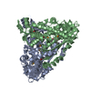
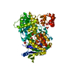
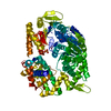
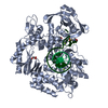
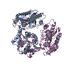
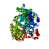
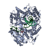
Crystal structure of the nucleotide binding domain FbpC complexed with ATP
Components
Fe(3+) ions import ATP-binding protein fbpC
Keywords
HYDROLASE / Nucleotide Binding Domain / ABC Motor Domain / Ferric Iron Transport / ATP-binding / Cell inner membrane / Cell membrane / Ion transport / Iron / Iron transport / Membrane / Nucleotide-binding / Transport
Function / homology
Function and homology information
ABC-type Fe3+ transporter / ABC-type ferric iron transporter activity / iron import into cell / ATP hydrolysis activity / ATP binding / plasma membrane Similarity search - Function
Mass: 18.015 Da / Num. of mol.: 522 / Source method: isolated from a natural source / Formula: H2O
Sequence details
AUTHORS STATE THAT THESE SEQUENCE CONFLICTS ARE DUE TO THIS GENE HAVING BEEN ISOLATED FROM A ...AUTHORS STATE THAT THESE SEQUENCE CONFLICTS ARE DUE TO THIS GENE HAVING BEEN ISOLATED FROM A CLINICAL ISOLATE OF N. GONORRHEA RATHER THAN THE ONE USED FOR THE SEQUENCING PROJECT. THESE MUTATIONS WERE PRESENT IN THE CLINICAL ISOLATE BUT ARE NOT IN ANY FUNCTIONALLY SIGNIFICANT AREAS OF THE PROTEIN.
-
Experimental details
-
Experiment
Experiment
Method: X-RAY DIFFRACTION / Number of used crystals: 1
-
Sample preparation
Crystal
Density Matthews: 2.38 Å3/Da / Density % sol: 48.4 %
Crystal grow
Temperature: 298 K / Method: vapor diffusion, sitting drop / pH: 6.5 Details: 20% PEG 6000, 0.1M MES, 0.2M CaCl2, pH 6.5, VAPOR DIFFUSION, SITTING DROP, temperature 298K
In the structure databanks used in Yorodumi, some data are registered as the other names, "COVID-19 virus" and "2019-nCoV". Here are the details of the virus and the list of structure data.
Jan 31, 2019. EMDB accession codes are about to change! (news from PDBe EMDB page)
EMDB accession codes are about to change! (news from PDBe EMDB page)
The allocation of 4 digits for EMDB accession codes will soon come to an end. Whilst these codes will remain in use, new EMDB accession codes will include an additional digit and will expand incrementally as the available range of codes is exhausted. The current 4-digit format prefixed with “EMD-” (i.e. EMD-XXXX) will advance to a 5-digit format (i.e. EMD-XXXXX), and so on. It is currently estimated that the 4-digit codes will be depleted around Spring 2019, at which point the 5-digit format will come into force.
The EM Navigator/Yorodumi systems omit the EMD- prefix.
Related info.:Q: What is EMD? / ID/Accession-code notation in Yorodumi/EM Navigator
Yorodumi is a browser for structure data from EMDB, PDB, SASBDB, etc.
This page is also the successor to EM Navigator detail page, and also detail information page/front-end page for Omokage search.
The word "yorodu" (or yorozu) is an old Japanese word meaning "ten thousand". "mi" (miru) is to see.
Related info.:EMDB / PDB / SASBDB / Comparison of 3 databanks / Yorodumi Search / Aug 31, 2016. New EM Navigator & Yorodumi / Yorodumi Papers / Jmol/JSmol / Function and homology information / Changes in new EM Navigator and Yorodumi
 Movie
Movie Controller

 Yorodumi
 Open data
 Basic information
 Components
 Keywords
 HYDROLASE / Nucleotide Binding Domain / ABC Motor Domain / Ferric Iron Transport / ATP-binding / Cell inner membrane /
  Function and homology information
 Authors
 Citation
 Structure visualization
 Downloads & links
 Other downloads
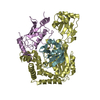


 PDBj
PDBj

 Assembly
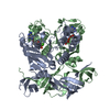

 Mass: 507.181 Da / Num. of mol.: 2 / Source method: obtained synthetically / Formula: C10H16N5O13P3 / Comment: ATP, energy-carrying molecule*YM
 Mass: 40.078 Da / Num. of mol.: 3 / Source method: obtained synthetically / Formula: Ca
 Mass: 18.015 Da / Num. of mol.: 522 / Source method: isolated from a natural source / Formula: H2O
 Sample preparation
 / Beamline: ID23-1 / Wavelength: 0.9797 Å
 Processing