 Movie
Movie Controller

[English] 日本語
 Yorodumi
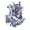
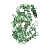
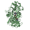
- PDB-3fim: Crystal structure of aryl-alcohol-oxidase from Pleurotus eryingii -
+ Open data

- Basic information
| Entry | Database: PDB / ID: 3fim | ||||||
|---|---|---|---|---|---|---|---|
| Title | Crystal structure of aryl-alcohol-oxidase from Pleurotus eryingii | ||||||
 Components Components | Aryl-alcohol oxidase | ||||||
 Keywords Keywords | OXIDOREDUCTASE / AAO / lignin degradation / Pleurotus eryngii / Flavoprotein | ||||||
| Function / homology |  Function and homology informationaryl-alcohol oxidase / aryl-alcohol oxidase activity / oxidoreductase activity, acting on paired donors, with incorporation or reduction of molecular oxygen / flavin adenine dinucleotide binding Function and homology informationaryl-alcohol oxidase / aryl-alcohol oxidase activity / oxidoreductase activity, acting on paired donors, with incorporation or reduction of molecular oxygen / flavin adenine dinucleotide bindingSimilarity search - Function | ||||||
| Biological species |  Pleurotus eryngii (fungus) Pleurotus eryngii (fungus) | ||||||
| Method | X-RAY DIFFRACTION / SYNCHROTRON / SAD / Resolution: 2.55 Å | ||||||
 Authors Authors | Fernandez, I.S. | ||||||
 Citation Citation | Journal: Acta Crystallogr.,Sect.D / Year: 2009 Title: Novel structural features in the GMC family of oxidoreductases revealed by the crystal structure of fungal aryl-alcohol oxidase Authors: Fernandez, I.S. / Ruiz-Duenas, F.J. / Santillana, E. / Ferreira, P. / Martinez, M.J. / Martinez, A.T. / Romero, A. | ||||||
| History |
|
- Structure visualization
| Structure viewer | Molecule: MolmilJmol/JSmol |
|---|
- Downloads & links
-Download
| PDBx/mmCIF format | 3fim.cif.gz | 123.1 KB | Display | PDBx/mmCIF format |
|---|---|---|---|---|
| PDB format | pdb3fim.ent.gz | 99.9 KB | Display | PDB format |
| PDBx/mmJSON format | 3fim.json.gz | Tree view | PDBx/mmJSON format | |
| Others |  Other downloads Other downloads |
-Validation report
| Arichive directory | https://data.pdbj.org/pub/pdb/validation_reports/fi/3fimftp://data.pdbj.org/pub/pdb/validation_reports/fi/3fim | HTTPS FTP |
|---|
-Related structure data
| Similar structure data |
|---|


-Links
 PDBj
PDBj
- Assembly
| Deposited unit | 
| ||||||||
|---|---|---|---|---|---|---|---|---|---|
| 1 |
| ||||||||
| Unit cell |
|
-Components
| #1: Protein | Mass: 60973.609 Da / Num. of mol.: 1 Source method: isolated from a genetically manipulated source Source: (gene. exp.) Pleurotus eryngii (fungus) / Gene: aao / Plasmid: pFLAG1 / Production host:  Escherichia coli (E. coli) / References: UniProt: O94219, aryl-alcohol oxidase Escherichia coli (E. coli) / References: UniProt: O94219, aryl-alcohol oxidase | ||
|---|---|---|---|
| #2: Chemical | Flavin adenine dinucleotide  Mass: 785.550 Da / Num. of mol.: 1 / Source method: obtained synthetically / Formula: C27H33N9O15P2 / Comment: FAD*YM Mass: 785.550 Da / Num. of mol.: 1 / Source method: obtained synthetically / Formula: C27H33N9O15P2 / Comment: FAD*YM#3: Water | ChemComp-HOH / | Water Mass: 18.015 Da / Num. of mol.: 225 / Source method: isolated from a natural source / Formula: H2O Mass: 18.015 Da / Num. of mol.: 225 / Source method: isolated from a natural source / Formula: H2O |
-Experimental details
-Experiment
| Experiment | Method: X-RAY DIFFRACTION / Number of used crystals: 2 |
|---|
- Sample preparation
| Crystal | Density Matthews: 6.17 Å3/Da / Density % sol: 80.08 % |
|---|---|
| Crystal grow | Temperature: 295 K / Method: vapor diffusion, sitting drop / pH: 7.4 Details: 1M lithium sulfate, 0.1M Bis-Tris propane, pH7.4, VAPOR DIFFUSION, SITTING DROP, temperature 295K |
-Data collection
| Diffraction |
| |||||||||||||||
|---|---|---|---|---|---|---|---|---|---|---|---|---|---|---|---|---|
| Diffraction source |
| |||||||||||||||
| Detector |
| |||||||||||||||
| Radiation |
| |||||||||||||||
| Radiation wavelength |
| |||||||||||||||
| Reflection | Resolution: 2.39→37.7 Å / Num. obs: 56141 / % possible obs: 97.7 % / Observed criterion σ(I): 2 / Redundancy: 15 % / Biso Wilson estimate: 35.68 Å2 / Rmerge(I) obs: 0.102 | |||||||||||||||
| Reflection shell | Resolution: 2.39→37.7 Å / Redundancy: 7.6 % / Rmerge(I) obs: 0.464 / Mean I/σ(I) obs: 4 / % possible all: 88.8 |


- Processing
| Software |
| ||||||||||||||||||||||||||||||||||||||||||||||||||||||||||||||||||||||||||||||||||||||||||
|---|---|---|---|---|---|---|---|---|---|---|---|---|---|---|---|---|---|---|---|---|---|---|---|---|---|---|---|---|---|---|---|---|---|---|---|---|---|---|---|---|---|---|---|---|---|---|---|---|---|---|---|---|---|---|---|---|---|---|---|---|---|---|---|---|---|---|---|---|---|---|---|---|---|---|---|---|---|---|---|---|---|---|---|---|---|---|---|---|---|---|---|
| Refinement | Method to determine structure: SAD / Resolution: 2.55→20.04 Å / Cor.coef. Fo:Fc: 0.96 / Cor.coef. Fo:Fc free: 0.944 / SU B: 8.365 / SU ML: 0.101 / Cross valid method: THROUGHOUT / ESU R: 0.148 / ESU R Free: 0.145 / Stereochemistry target values: MAXIMUM LIKELIHOOD / Details: HYDROGENS HAVE BEEN ADDED IN THE RIDING POSITIONS
| ||||||||||||||||||||||||||||||||||||||||||||||||||||||||||||||||||||||||||||||||||||||||||
| Solvent computation | Ion probe radii: 0.8 Å / Shrinkage radii: 0.8 Å / VDW probe radii: 1.2 Å / Solvent model: MASK | ||||||||||||||||||||||||||||||||||||||||||||||||||||||||||||||||||||||||||||||||||||||||||
| Displacement parameters | Biso mean: 35.683 Å2
| ||||||||||||||||||||||||||||||||||||||||||||||||||||||||||||||||||||||||||||||||||||||||||
| Refinement step | Cycle: LAST / Resolution: 2.55→20.04 Å
| ||||||||||||||||||||||||||||||||||||||||||||||||||||||||||||||||||||||||||||||||||||||||||
| Refine LS restraints |
| ||||||||||||||||||||||||||||||||||||||||||||||||||||||||||||||||||||||||||||||||||||||||||
| LS refinement shell | Resolution: 2.392→2.454 Å / Total num. of bins used: 20
| ||||||||||||||||||||||||||||||||||||||||||||||||||||||||||||||||||||||||||||||||||||||||||
| Refinement TLS params. | Method: refined / Origin x: -21.244 Å / Origin y: 67.828 Å / Origin z: -10.402 Å
| ||||||||||||||||||||||||||||||||||||||||||||||||||||||||||||||||||||||||||||||||||||||||||
| Refinement TLS group |
|
