 Movie
Movie Controller

[English] 日本語
 Yorodumi
- PDB-3fhp: A neutron crystallographic analysis of a porcine 2Zn insulin at 2... -
+ Open data

- Basic information
| Entry | Database: PDB / ID: 3fhp | ||||||
|---|---|---|---|---|---|---|---|
| Title | A neutron crystallographic analysis of a porcine 2Zn insulin at 2.0 A resolution | ||||||
 Components Components | (Insulin ) x 2 ) x 2 | ||||||
 Keywords Keywords | HORMONE / 2Zn insulin / neutron crystallography / protonation / H/D exchange / Carbohydrate metabolism / Cleavage on pair of basic residues / Glucose metabolism / Secreted | ||||||
| Function / homology |  Function and homology information Function and homology informationInsulin processing / IRS activation / Signal attenuation / Insulin receptor signalling cascade / Signaling by Insulin receptor / Synthesis, secretion, and deacylation of Ghrelin / PI5P, PP2A and IER3 Regulate PI3K/AKT Signaling / Insulin receptor recycling / glycoprotein biosynthetic process / response to L-arginine ...Insulin processing / IRS activation / Signal attenuation / Insulin receptor signalling cascade / Signaling by Insulin receptor / Synthesis, secretion, and deacylation of Ghrelin / PI5P, PP2A and IER3 Regulate PI3K/AKT Signaling / Insulin receptor recycling / glycoprotein biosynthetic process / response to L-arginine / positive regulation of lipoprotein lipase activity / lactate biosynthetic process / lipoprotein biosynthetic process / positive regulation of fatty acid biosynthetic process / positive regulation of glucose metabolic process / COPI-mediated anterograde transport / lipid biosynthetic process / negative regulation of glycogen catabolic process / regulation of cellular amino acid metabolic process / nitric oxide-cGMP-mediated signaling / negative regulation of fatty acid metabolic process / negative regulation of feeding behavior / positive regulation of respiratory burst / positive regulation of dendritic spine maintenance / alpha-beta T cell activation / negative regulation of acute inflammatory response / negative regulation of respiratory burst involved in inflammatory response / negative regulation of protein secretion / fatty acid homeostasis / positive regulation of glycogen biosynthetic process / positive regulation of DNA replication / negative regulation of gluconeogenesis / positive regulation of nitric oxide mediated signal transduction / regulation of protein localization to plasma membrane / negative regulation of lipid catabolic process / negative regulation of reactive oxygen species biosynthetic process / positive regulation of insulin receptor signaling pathway / positive regulation of protein autophosphorylation / insulin-like growth factor receptor binding / neuron projection maintenance / positive regulation of glycolytic process / positive regulation of mitotic nuclear division / positive regulation of cytokine production / acute-phase response / positive regulation of protein secretion / positive regulation of glucose import / negative regulation of proteolysis / wound healing / insulin receptor binding / negative regulation of protein catabolic process / hormone activity / vasodilation / positive regulation of protein localization to nucleus / glucose metabolic process / glucose homeostasis / insulin receptor signaling pathway / protease binding / positive regulation of MAPK cascade / positive regulation of phosphatidylinositol 3-kinase/protein kinase B signal transduction / positive regulation of cell migration / G protein-coupled receptor signaling pathway / negative regulation of gene expression / positive regulation of cell population proliferation / extracellular space / identical protein bindingSimilarity search - Function | ||||||
| Biological species |  Sus scrofa (pig) Sus scrofa (pig) | ||||||
| Method | NEUTRON DIFFRACTION / NUCLEAR REACTOR / MOLECULAR REPLACEMENT / Resolution: 2 Å | ||||||
 Authors Authors | Iwai, W. / Kurihara, K. / Yamada, T. / Kobayashi, Y. / Ohnishi, Y. / Tanaka, I. / Takahashi, H. / Niimura, N. | ||||||
 Citation Citation | Journal: Acta Crystallogr.,Sect.D / Year: 2009 Title: A neutron crystallographic analysis of T6 porcine insulin at 2.1 A resolution Authors: Iwai, W. / Yamada, T. / Kurihara, K. / Ohnishi, Y. / Kobayashi, Y. / Tanaka, I. / Takahashi, H. / Kuroki, R. / Tamada, T. / Niimura, N. | ||||||
| History |
|
- Structure visualization
| Structure viewer | Molecule: MolmilJmol/JSmol |
|---|
- Downloads & links
-Download
| PDBx/mmCIF format | 3fhp.cif.gz | 56.7 KB | Display | PDBx/mmCIF format |
|---|---|---|---|---|
| PDB format | pdb3fhp.ent.gz | 42.9 KB | Display | PDB format |
| PDBx/mmJSON format | 3fhp.json.gz | Tree view | PDBx/mmJSON format | |
| Others |  Other downloads Other downloads |
-Validation report
| Arichive directory | https://data.pdbj.org/pub/pdb/validation_reports/fh/3fhpftp://data.pdbj.org/pub/pdb/validation_reports/fh/3fhp | HTTPS FTP |
|---|
-Related structure data
| Related structure data | 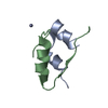 4insS S: Starting model for refinement |
|---|---|
| Similar structure data |
-Links
 PDBj
PDBj


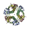
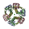
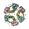
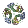
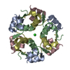
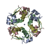
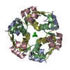
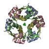
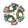
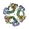
- Assembly
| Deposited unit | 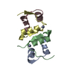
| |||||||||||||||
|---|---|---|---|---|---|---|---|---|---|---|---|---|---|---|---|---|
| 1 | 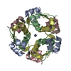
| |||||||||||||||
| Unit cell |
| |||||||||||||||
| Components on special symmetry positions |
|
-Components
| #1: Protein/peptide | / Insulin A chain Mass: 2383.698 Da / Num. of mol.: 2 / Fragment: UNP residues 88-108 Source method: isolated from a genetically manipulated source Source: (gene. exp.) Sus scrofa (pig) / Gene: INS / Production host:  Escherichia coli (E. coli) / References: UniProt: P01315 Escherichia coli (E. coli) / References: UniProt: P01315#2: Protein/peptide | / Insulin B chainMass: 3403.927 Da / Num. of mol.: 2 / Fragment: UNP residues 25-54 Source method: isolated from a genetically manipulated source Source: (gene. exp.) Sus scrofa (pig) / Gene: INS / Production host: Escherichia coli (E. coli) / References: UniProt: P01315#3: Chemical |   Mass: 65.409 Da / Num. of mol.: 2 / Source method: obtained synthetically / Formula: Zn Mass: 65.409 Da / Num. of mol.: 2 / Source method: obtained synthetically / Formula: Zn#4: Chemical | ChemComp-DOD / | Heavy water  Mass: 18.015 Da / Num. of mol.: 89 / Source method: isolated from a natural source / Formula: D2O Mass: 18.015 Da / Num. of mol.: 89 / Source method: isolated from a natural source / Formula: D2O |
|---|
-Experimental details
-Experiment
| Experiment | Method: NEUTRON DIFFRACTION / Number of used crystals: 1 |
|---|
- Sample preparation
| Crystal grow | Temperature: 293 K / Method: liquid diffusion / pH: 6.3 Details: 2.3mg/ml insulin, 50mM sodium citrate, 6mM zinc acetate, 17% acetone, pH6.3, LIQUID DIFFUSION, temperature 293K |
|---|
-Data collection
| Diffraction | Mean temperature: 298 K |
|---|---|
| Diffraction source | Source: NUCLEAR REACTOR / Site: JRR-3M  / Beamline: 1G-B / Wavelength: 2.6 Å / Beamline: 1G-B / Wavelength: 2.6 Å |
| Detector | Type: MACSCIENCE / Detector: IMAGE PLATE / Date: May 8, 2006 |
| Radiation | Monochromator: ELLASTICALLY BENT SILICON / Protocol: SINGLE WAVELENGTH / Monochromatic (M) / Laue (L): M / Scattering type: neutron |
| Radiation wavelength | Wavelength: 2.6 Å / Relative weight: 1 |
| Reflection | Resolution: 2→80 Å / Num. all: 4824 / Num. obs: 4824 / % possible obs: 23.2 % / Redundancy: 2.2 % / Biso Wilson estimate: 8.2 Å2 / Rmerge(I) obs: 0.147 |
| Reflection shell | Resolution: 2→2.07 Å / Redundancy: 2.2 % / Rmerge(I) obs: 0.393 / Num. unique all: 4824 / % possible all: 81.3 |
- Processing
| Software |
| ||||||||||||||||||||||||||||||||||||||||||||||||||||||||||||||||||||||||||||||||
|---|---|---|---|---|---|---|---|---|---|---|---|---|---|---|---|---|---|---|---|---|---|---|---|---|---|---|---|---|---|---|---|---|---|---|---|---|---|---|---|---|---|---|---|---|---|---|---|---|---|---|---|---|---|---|---|---|---|---|---|---|---|---|---|---|---|---|---|---|---|---|---|---|---|---|---|---|---|---|---|---|---|
| Refinement | Method to determine structure: MOLECULAR REPLACEMENT Starting model: PDB ENTRY 4INS Resolution: 2→41.43 Å / Rfactor Rfree error: 0.017 / Data cutoff high absF: 484314.36 / Data cutoff low absF: 0 / Isotropic thermal model: RESTRAINED / Cross valid method: THROUGHOUT / σ(F): 3
| ||||||||||||||||||||||||||||||||||||||||||||||||||||||||||||||||||||||||||||||||
| Solvent computation | Solvent model: FLAT MODEL / Bsol: 24.8544 Å2 / ksol: 0.06 e/Å3 | ||||||||||||||||||||||||||||||||||||||||||||||||||||||||||||||||||||||||||||||||
| Displacement parameters | Biso mean: 23.8 Å2
| ||||||||||||||||||||||||||||||||||||||||||||||||||||||||||||||||||||||||||||||||
| Refine analyze |
| ||||||||||||||||||||||||||||||||||||||||||||||||||||||||||||||||||||||||||||||||
| Refinement step | Cycle: LAST / Resolution: 2→41.43 Å
| ||||||||||||||||||||||||||||||||||||||||||||||||||||||||||||||||||||||||||||||||
| Refine LS restraints |
| ||||||||||||||||||||||||||||||||||||||||||||||||||||||||||||||||||||||||||||||||
| LS refinement shell | Resolution: 2→2.13 Å / Rfactor Rfree error: 0.075 / Total num. of bins used: 6
| ||||||||||||||||||||||||||||||||||||||||||||||||||||||||||||||||||||||||||||||||
| Xplor file |
|
