 Movie
Movie Controller

[English] 日本語
 Yorodumi
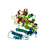
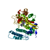
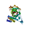
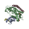
- PDB-3fhb: Human poly(ADP-ribose) polymerase 3, catalytic fragment in comple... -
+ Open data

- Basic information
| Entry | Database: PDB / ID: 3fhb | |||||||||
|---|---|---|---|---|---|---|---|---|---|---|
| Title | Human poly(ADP-ribose) polymerase 3, catalytic fragment in complex with an inhibitor 3-aminobenzoic acid | |||||||||
 Components Components | Poly [ADP-ribose] polymerase 3 | |||||||||
 Keywords Keywords |  TRANSFERASE / enzyme-inhibitor complex / catalytic fragment / Structural Genomics / Structural Genomics Consortium / SGC / Alternative splicing / Glycosyltransferase / NAD / Nucleus TRANSFERASE / enzyme-inhibitor complex / catalytic fragment / Structural Genomics / Structural Genomics Consortium / SGC / Alternative splicing / Glycosyltransferase / NAD / Nucleus | |||||||||
| Function / homology |  Function and homology information Function and homology informationnegative regulation of isotype switching / negative regulation of telomerase RNA reverse transcriptase activity / NAD+- protein-lysine ADP-ribosyltransferase activity / positive regulation of DNA ligation / NAD DNA ADP-ribosyltransferase activity / NAD+- protein-aspartate ADP-ribosyltransferase activity / NAD+-protein-glutamate ADP-ribosyltransferase activity / DNA ADP-ribosylation / protein localization to site of double-strand break / protein auto-ADP-ribosylation ...negative regulation of isotype switching / negative regulation of telomerase RNA reverse transcriptase activity / NAD+- protein-lysine ADP-ribosyltransferase activity / positive regulation of DNA ligation / NAD DNA ADP-ribosyltransferase activity / NAD+- protein-aspartate ADP-ribosyltransferase activity / NAD+-protein-glutamate ADP-ribosyltransferase activity / DNA ADP-ribosylation / protein localization to site of double-strand break / protein auto-ADP-ribosylation / intercellular bridge / positive regulation of double-strand break repair via nonhomologous end joining / NAD+-protein ADP-ribosyltransferase activity / NAD+ ADP-ribosyltransferase activity / Transferases; Glycosyltransferases; Pentosyltransferases / catalytic activity / regulation of mitotic spindle organization / telomere maintenance / centriole / nucleotidyltransferase activity / double-strand break repair / site of double-strand break / nuclear body / centrosome / nucleolus / nucleoplasm / cytoplasmSimilarity search - Function | |||||||||
| Biological species |  Homo sapiens (human) Homo sapiens (human) | |||||||||
| Method | X-RAY DIFFRACTION / SYNCHROTRON / MOLECULAR REPLACEMENT / Resolution: 2.3 Å | |||||||||
 Authors Authors | Lehtio, L. / Karlberg, T. / Arrowsmith, C.H. / Berglund, H. / Busam, R. / Collins, R. / Dahlgren, L.G. / Edwards, A.M. / Flodin, S. / Flores, A. ...Lehtio, L. / Karlberg, T. / Arrowsmith, C.H. / Berglund, H. / Busam, R. / Collins, R. / Dahlgren, L.G. / Edwards, A.M. / Flodin, S. / Flores, A. / Graslund, S. / Hammarstrom, M. / Hallberg, B.M. / Johansson, I. / Kotenyova, T. / Moche, M. / Nordlund, P. / Nyman, T. / Ogg, D. / Persson, C. / Sagemark, J. / Schueler, H. / Stenmark, P. / Sundstrom, M. / Thorsell, A.G. / Van Den Berg, S. / Weigelt, J. / Holmberg-Schiavone, L. / Structural Genomics Consortium (SGC) | |||||||||
 Citation Citation | Journal: J.Med.Chem. / Year: 2009 Title: Structural basis for inhibitor specificity in human poly(ADP-ribose) polymerase-3. Authors: Lehtio, L. / Jemth, A.S. / Collins, R. / Loseva, O. / Johansson, A. / Markova, N. / Hammarstrom, M. / Flores, A. / Holmberg-Schiavone, L. / Weigelt, J. / Helleday, T. / Schuler, H. / Karlberg, T. | |||||||||
| History |
|
- Structure visualization
| Structure viewer | Molecule: MolmilJmol/JSmol |
|---|
- Downloads & links
-Download
| PDBx/mmCIF format | 3fhb.cif.gz | 85.2 KB | Display | PDBx/mmCIF format |
|---|---|---|---|---|
| PDB format | pdb3fhb.ent.gz | 62.8 KB | Display | PDB format |
| PDBx/mmJSON format | 3fhb.json.gz | Tree view | PDBx/mmJSON format | |
| Others |  Other downloads Other downloads |
-Validation report
| Arichive directory | https://data.pdbj.org/pub/pdb/validation_reports/fh/3fhbftp://data.pdbj.org/pub/pdb/validation_reports/fh/3fhb | HTTPS FTP |
|---|
-Related structure data


-Links
 PDBj
PDBj- Assembly
| Deposited unit | 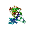
| ||||||||
|---|---|---|---|---|---|---|---|---|---|
| 1 |
| ||||||||
| Unit cell |
|
-Components
| #1: Protein | Mass: 39752.074 Da / Num. of mol.: 1 / Fragment: Catalytic fragment: UNP residues 178-532 Source method: isolated from a genetically manipulated source Source: (gene. exp.) Homo sapiens (human) / Gene: PARP3, ADPRT3, ADPRTL3 / Plasmid: pNIC-Bsa4 / Production host:  Escherichia coli (E. coli) / Strain (production host): BL21(DE3) / References: UniProt: Q9Y6F1, NAD+ ADP-ribosyltransferase Escherichia coli (E. coli) / Strain (production host): BL21(DE3) / References: UniProt: Q9Y6F1, NAD+ ADP-ribosyltransferase |
|---|---|
| #2: Chemical | ChemComp-GAB / 3-Aminobenzoic acid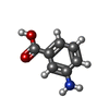  Mass: 137.136 Da / Num. of mol.: 1 / Source method: obtained synthetically / Formula: C7H7NO2 Mass: 137.136 Da / Num. of mol.: 1 / Source method: obtained synthetically / Formula: C7H7NO2 |
| #3: Water | ChemComp-HOH / Water Mass: 18.015 Da / Num. of mol.: 144 / Source method: isolated from a natural source / Formula: H2O Mass: 18.015 Da / Num. of mol.: 144 / Source method: isolated from a natural source / Formula: H2O |
-Experimental details
-Experiment
| Experiment | Method: X-RAY DIFFRACTION / Number of used crystals: 1 |
|---|
- Sample preparation
| Crystal | Density Matthews: 2.05 Å3/Da / Density % sol: 40.01 % |
|---|---|
| Crystal grow | Temperature: 298 K / Method: vapor diffusion / pH: 7 Details: 1.8 M DL-Malic acid, 0.1 M Bis-Tris propane - seeding, pH 7.0, VAPOR DIFFUSION, temperature 298K |
-Data collection
| Diffraction | Mean temperature: 100 K |
|---|---|
| Diffraction source | Source: SYNCHROTRON / Site: ESRF  / Beamline: BM14 / Wavelength: 0.98 Å / Beamline: BM14 / Wavelength: 0.98 Å |
| Detector | Type: MARMOSAIC 225 mm CCD / Detector: CCD / Date: Sep 14, 2006 |
| Radiation | Monochromator: Si(111) / Protocol: SINGLE WAVELENGTH / Monochromatic (M) / Laue (L): M / Scattering type: x-ray |
| Radiation wavelength | Wavelength: 0.98 Å / Relative weight: 1 |
| Reflection | Resolution: 2.3→20 Å / Num. all: 14283 / Num. obs: 14283 / % possible obs: 98.5 % / Observed criterion σ(F): 0 / Observed criterion σ(I): 0 / Redundancy: 3.7 % / Biso Wilson estimate: 20.8 Å2 / Rmerge(I) obs: 0.156 / Net I/σ(I): 8.73 |
| Reflection shell | Resolution: 2.3→2.4 Å / Redundancy: 3.65 % / Rmerge(I) obs: 0.48 / Mean I/σ(I) obs: 2.73 / Num. unique all: 1666 / % possible all: 96.3 |
- Processing
| Software |
| ||||||||||||||||||||||||||||||||||||||||||||||||||||||||||||||||||||||||||||||||||||||||||||||||||||||||||||||||||||||||||||||||||||||||||||||||||||||||||||||||||||||||||
|---|---|---|---|---|---|---|---|---|---|---|---|---|---|---|---|---|---|---|---|---|---|---|---|---|---|---|---|---|---|---|---|---|---|---|---|---|---|---|---|---|---|---|---|---|---|---|---|---|---|---|---|---|---|---|---|---|---|---|---|---|---|---|---|---|---|---|---|---|---|---|---|---|---|---|---|---|---|---|---|---|---|---|---|---|---|---|---|---|---|---|---|---|---|---|---|---|---|---|---|---|---|---|---|---|---|---|---|---|---|---|---|---|---|---|---|---|---|---|---|---|---|---|---|---|---|---|---|---|---|---|---|---|---|---|---|---|---|---|---|---|---|---|---|---|---|---|---|---|---|---|---|---|---|---|---|---|---|---|---|---|---|---|---|---|---|---|---|---|---|---|---|
| Refinement | Method to determine structure: MOLECULAR REPLACEMENT / Resolution: 2.3→19.17 Å / Cor.coef. Fo:Fc: 0.939 / Cor.coef. Fo:Fc free: 0.884 / SU B: 8.352 / SU ML: 0.197 / Cross valid method: THROUGHOUT / σ(F): 0 / ESU R: 0.52 / ESU R Free: 0.277 / Stereochemistry target values: MAXIMUM LIKELIHOOD / Details: HYDROGENS HAVE BEEN ADDED IN THE RIDING POSITIONS
| ||||||||||||||||||||||||||||||||||||||||||||||||||||||||||||||||||||||||||||||||||||||||||||||||||||||||||||||||||||||||||||||||||||||||||||||||||||||||||||||||||||||||||
| Solvent computation | Ion probe radii: 0.8 Å / Shrinkage radii: 0.8 Å / VDW probe radii: 1.2 Å / Solvent model: MASK | ||||||||||||||||||||||||||||||||||||||||||||||||||||||||||||||||||||||||||||||||||||||||||||||||||||||||||||||||||||||||||||||||||||||||||||||||||||||||||||||||||||||||||
| Displacement parameters | Biso mean: 16.759 Å2
| ||||||||||||||||||||||||||||||||||||||||||||||||||||||||||||||||||||||||||||||||||||||||||||||||||||||||||||||||||||||||||||||||||||||||||||||||||||||||||||||||||||||||||
| Refinement step | Cycle: LAST / Resolution: 2.3→19.17 Å
| ||||||||||||||||||||||||||||||||||||||||||||||||||||||||||||||||||||||||||||||||||||||||||||||||||||||||||||||||||||||||||||||||||||||||||||||||||||||||||||||||||||||||||
| Refine LS restraints |
| ||||||||||||||||||||||||||||||||||||||||||||||||||||||||||||||||||||||||||||||||||||||||||||||||||||||||||||||||||||||||||||||||||||||||||||||||||||||||||||||||||||||||||
| LS refinement shell | Resolution: 2.3→2.36 Å / Total num. of bins used: 20
|
