 Movie
Movie Controller

[English] 日本語
 Yorodumi
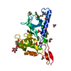
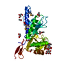
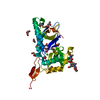
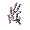
- PDB-3f5a: Crystal structure of Toxoplasma gondii micronemal protein 1 bound... -
+ Open data

- Basic information
| Entry | Database: PDB / ID: 3f5a | |||||||||
|---|---|---|---|---|---|---|---|---|---|---|
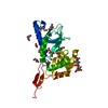
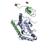
| Title | Crystal structure of Toxoplasma gondii micronemal protein 1 bound to 3'SiaLacNAc1-3 | |||||||||
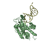
 Components Components | Micronemal protein 1 | |||||||||
 Keywords Keywords |  CELL ADHESION / cellular adhesion / micronemal protein / toxoplasma gondii / carbohydrate / fluorine / Cytoplasmic vesicle / Lectin / Virulence CELL ADHESION / cellular adhesion / micronemal protein / toxoplasma gondii / carbohydrate / fluorine / Cytoplasmic vesicle / Lectin / Virulence | |||||||||
| Function / homology |  Function and homology information Function and homology information | |||||||||
| Biological species |  Toxoplasma gondii (eukaryote) Toxoplasma gondii (eukaryote) | |||||||||
| Method | X-RAY DIFFRACTION / SYNCHROTRON / MOLECULAR REPLACEMENT / Resolution: 2 Å | |||||||||
 Authors Authors | Garnett, J.A. / Liu, Y. / Feizi, T. / Matthews, S.J. | |||||||||
 Citation Citation | Journal: Protein Sci. / Year: 2009 Title: Detailed insights from microarray and crystallographic studies into carbohydrate recognition by microneme protein 1 (MIC1) of Toxoplasma gondii. Authors: Garnett, J.A. / Liu, Y. / Leon, E. / Allman, S.A. / Friedrich, N. / Saouros, S. / Curry, S. / Soldati-Favre, D. / Davis, B.G. / Feizi, T. / Matthews, S. #1: Journal: Embo J. / Year: 2007Title: Atomic resolution insight into host cell recognition by Toxoplasma gondii. Authors: Blumenschein, T.M. / Friedrich, N. / Childs, R.A. / Saouros, S. / Carpenter, E.P. / Campanero-Rhodes, M.A. / Simpson, P. / Chai, W. / Koutroukides, T. / Blackman, M.J. / Feizi, T. / Soldati- ...Authors: Blumenschein, T.M. / Friedrich, N. / Childs, R.A. / Saouros, S. / Carpenter, E.P. / Campanero-Rhodes, M.A. / Simpson, P. / Chai, W. / Koutroukides, T. / Blackman, M.J. / Feizi, T. / Soldati-Favre, D. / Matthews, S. | |||||||||
| History |
|
- Structure visualization
| Structure viewer | Molecule: MolmilJmol/JSmol |
|---|
- Downloads & links
-Download
| PDBx/mmCIF format | 3f5a.cif.gz | 68.4 KB | Display | PDBx/mmCIF format |
|---|---|---|---|---|
| PDB format | pdb3f5a.ent.gz | 47.8 KB | Display | PDB format |
| PDBx/mmJSON format | 3f5a.json.gz | Tree view | PDBx/mmJSON format | |
| Others |  Other downloads Other downloads |
-Validation report
| Arichive directory | https://data.pdbj.org/pub/pdb/validation_reports/f5/3f5aftp://data.pdbj.org/pub/pdb/validation_reports/f5/3f5a | HTTPS FTP |
|---|
-Related structure data
| Related structure data |  3f53C  3f5eC 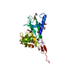 2jh1S S: Starting model for refinement C: citing same article ( |
|---|---|
| Similar structure data |


-Links
 PDBj
PDBj- Assembly
| Deposited unit | 
| ||||||||||||
|---|---|---|---|---|---|---|---|---|---|---|---|---|---|
| 1 |
| ||||||||||||
| Unit cell |
| ||||||||||||
| Components on special symmetry positions |
|
-Components
-Protein / Sugars , 2 types, 2 molecules A
| #1: Protein | Mass: 27266.010 Da / Num. of mol.: 1 / Fragment: N-terminal domain: Residues 17-262 Source method: isolated from a genetically manipulated source Source: (gene. exp.) Toxoplasma gondii (eukaryote) / Strain: RH / Gene: MIC1 / Plasmid: pET / Production host:  Escherichia coli (E. coli) / Strain (production host): origami / References: UniProt: O00834 Escherichia coli (E. coli) / Strain (production host): origami / References: UniProt: O00834 |
|---|---|
| #2: Polysaccharide | N-acetyl-alpha-neuraminic acid-(2-3)-beta-D-galactopyranose-(1-3)-2-acetamido-2-deoxy-beta-D-glucopyranose / Mass: 674.604 Da / Num. of mol.: 1 Source method: isolated from a genetically manipulated source |
-Non-polymers , 4 types, 203 molecules 


| #3: Chemical | ChemComp-CL / Chloride Mass: 35.453 Da / Num. of mol.: 1 / Source method: obtained synthetically / Formula: Cl Mass: 35.453 Da / Num. of mol.: 1 / Source method: obtained synthetically / Formula: Cl | ||||
|---|---|---|---|---|---|
| #4: Chemical | ChemComp-ACT / Acetate Mass: 59.044 Da / Num. of mol.: 8 / Source method: obtained synthetically / Formula: C2H3O2 Mass: 59.044 Da / Num. of mol.: 8 / Source method: obtained synthetically / Formula: C2H3O2#5: Chemical | ChemComp-GOL / Glycerol Mass: 92.094 Da / Num. of mol.: 5 / Source method: obtained synthetically / Formula: C3H8O3 Mass: 92.094 Da / Num. of mol.: 5 / Source method: obtained synthetically / Formula: C3H8O3#6: Water | ChemComp-HOH / | WaterMass: 18.015 Da / Num. of mol.: 189 / Source method: isolated from a natural source / Formula: H2O |
-Experimental details
-Experiment
| Experiment | Method: X-RAY DIFFRACTION / Number of used crystals: 1 |
|---|
- Sample preparation
| Crystal | Density Matthews: 3.46 Å3/Da / Density % sol: 64.47 % |
|---|---|
| Crystal grow | Temperature: 290 K / Method: vapor diffusion, hanging drop / pH: 7.6 Details: 3.6M Ammonium acetate, 100mM Bis/tris-propane, pH 7.6, VAPOR DIFFUSION, HANGING DROP, temperature 290K |
-Data collection
| Diffraction | Mean temperature: 100 K |
|---|---|
| Diffraction source | Source: SYNCHROTRON / Site: ESRF  / Beamline: BM14 / Wavelength: 0.934 Å / Beamline: BM14 / Wavelength: 0.934 Å |
| Detector | Type: ADSC QUANTUM 4 / Detector: CCD / Date: Sep 19, 2007 / Details: mirrors |
| Radiation | Protocol: SINGLE WAVELENGTH / Monochromatic (M) / Laue (L): M / Scattering type: x-ray |
| Radiation wavelength | Wavelength: 0.934 Å / Relative weight: 1 |
| Reflection | Resolution: 2→33.17 Å / Num. obs: 23609 / % possible obs: 99.3 % / Redundancy: 3.5 % / Biso Wilson estimate: 23.545 Å2 / Rsym value: 0.074 / Net I/σ(I): 11.2 |
| Reflection shell | Resolution: 2→2.11 Å / Redundancy: 3.6 % / Mean I/σ(I) obs: 4.7 / Num. unique all: 3837 / Rsym value: 0.2228 / % possible all: 100 |
- Processing
| Software |
| ||||||||||||||||||||||||||||||||||||||||||||||||||||||||||||||||||||||||||||||||||||||||||
|---|---|---|---|---|---|---|---|---|---|---|---|---|---|---|---|---|---|---|---|---|---|---|---|---|---|---|---|---|---|---|---|---|---|---|---|---|---|---|---|---|---|---|---|---|---|---|---|---|---|---|---|---|---|---|---|---|---|---|---|---|---|---|---|---|---|---|---|---|---|---|---|---|---|---|---|---|---|---|---|---|---|---|---|---|---|---|---|---|---|---|---|
| Refinement | Method to determine structure: MOLECULAR REPLACEMENT Starting model: PDB entry 2JH1 Resolution: 2→27.75 Å / Cor.coef. Fo:Fc: 0.96 / Cor.coef. Fo:Fc free: 0.949 / SU B: 4.514 / SU ML: 0.067 / Isotropic thermal model: TLS / Cross valid method: THROUGHOUT / ESU R: 0.126 / ESU R Free: 0.116 / Stereochemistry target values: MAXIMUM LIKELIHOOD / Details: HYDROGENS HAVE BEEN ADDED IN THE RIDING POSITIONS
| ||||||||||||||||||||||||||||||||||||||||||||||||||||||||||||||||||||||||||||||||||||||||||
| Solvent computation | Ion probe radii: 0.8 Å / Shrinkage radii: 0.8 Å / VDW probe radii: 1.2 Å / Solvent model: MASK | ||||||||||||||||||||||||||||||||||||||||||||||||||||||||||||||||||||||||||||||||||||||||||
| Displacement parameters | Biso mean: 21.913 Å2
| ||||||||||||||||||||||||||||||||||||||||||||||||||||||||||||||||||||||||||||||||||||||||||
| Refinement step | Cycle: LAST / Resolution: 2→27.75 Å
| ||||||||||||||||||||||||||||||||||||||||||||||||||||||||||||||||||||||||||||||||||||||||||
| Refine LS restraints |
| ||||||||||||||||||||||||||||||||||||||||||||||||||||||||||||||||||||||||||||||||||||||||||
| LS refinement shell | Resolution: 2→2.052 Å / Total num. of bins used: 20
|
